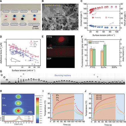
Fig. 2. Characterization of the fluid interfacing and light sensing.

(A) Image of droplets of water, ethanol, acetone, dichloromethane (DCM), silicone oil (PDMS), n-heptane, dimethylformamide (DMF), and ethyl acetate residing atop the translucent superomniphobic surface. (B) SEM image showing the fractal network of the superomniphobic surface. Inset shows the typical inverted structures. (C) Super-repellency toward various liquids. (D) Adhesive force is inversely proportional to the surface tension. Error bars denote SD of three independent measurements. (E) Liquid residue detected on diverse omniphobic surfaces by fluorescence imaging. (F) Fluorescence intensity and area fraction of the images in (E), showing the remarkably reduced liquid loss on the superomniphobic (SOP) surface. Error bars denote SD of three independent measurements. (G) Sequential images showing an n-heptane droplet (r0 ≈ 1 mm, We ≈ 20) bounces on the surface, exhibiting low adhesion toward organic liquids. Time interval between each snapshot is ~4 ms. (H) Infrared thermal imaging and the plot showing the temperature distribution on photothermal film upon 400-mW laser irradiation. (I) Thermal response of graphene-PDMS composite films with varying contents of graphene nanoplatelets to 400-mW laser irradiation. Blue and red shaded regions denote off and on states, respectively, of the 785-nm laser. (J) Thermal response of PDMS film containing 5 wt % graphene nanoplatelets to laser power. The solid lines are from theoretical analysis (see note S2 for details). Photo credit: Wei Li, The University of Hong Kong.
