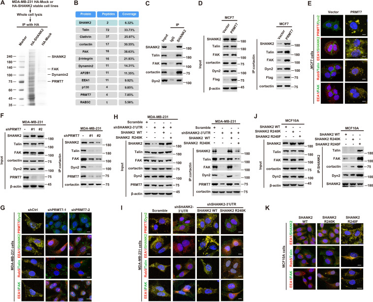
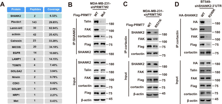
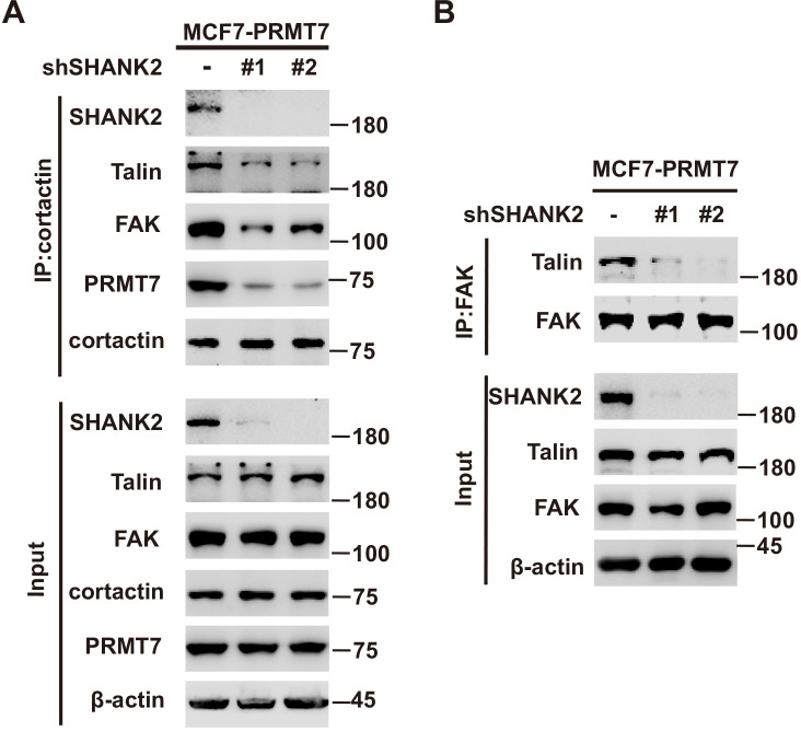
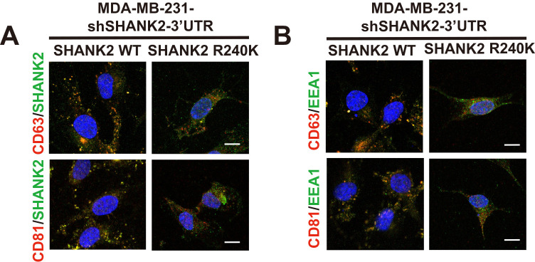
Figure 5. SHANK2 R240 methylation promotes FAK/dynamin2/talin complex co-localized with endosome.
(A) SHANK2 associated proteins from HA-SHANK2 expressing stable MDA-MB-231 cells were immunopurified with anti-HA (α-HA) affinity resins. The protein bands were analyzed by mass spectrometry. (B) Representative peptide fragments of SHANK2 associated proteins and peptide coverage of the indicated proteins are shown. (C) Endogenous talin, FAK, dynamin2, EEA1 and SHANK2 were IP from MDA-MB-231 cells, with indicated antibodies, and the binding of talin, FAK, dynamin2 and SHANK2 was examined by western blot. (D) MCF7 cells with expression of the indicated Flag-Vector or Flag-tagged PRMT7. Immunoprecipitation of cortactin with anti-cortactin antibody was performed. (E) Confocal images of PRMT7, dynamin2, SHANK2, EEA1, talin, Rab5, FAK, cortactin and DAPI staining in MCF7 cells with expression of the indicated Flag-Vector or Flag-tagged PRMT7. Scale bars, 10 µm. (F) MDA-MB-231cells with or without stable expression of the indicated PRMT7 shRNA or a control shRNA. Immunoprecipitation of cortactin with anti-cortactin antibody was performed. (G) confocal images of PRMT7, dynamin2, SHANK2, EEA1, talin, Rab5, FAK, cortactin and DAPI staining in MDA-MB-231cells with or without stable expression of the indicated PRMT7 shRNA or a control shRNA. Scale bars, 10 µm. (H) MDA-MB-231cells expressing SHANK2 shRNA with or without reconstituted expression of WT SHANK2 or SHANK2 R240K mutant. Immunoprecipitation of cortactin with anti-cortactin antibody was performed. (I) Confocal images of PRMT7, dynamin2, SHANK2, EEA1, talin, Rab5, FAK, cortactin and DAPI staining in MDA-MB-231cells expressing SHANK2 shRNA with or without reconstituted expression of WT SHANK2 or SHANK2 R240K mutant. Scale bars, 10 µm. (J) MCF10A cells with reconstituted expression of SHANK2 WT, SHANK2 R240K or SHANK2 R240F (arginine to phenylalanine) mutant. Immunoprecipitation of SHANK2 with anti-SHANK2 antibody was performed. (K) Confocal images of PRMT7, dynamin2, SHANK2, EEA1, talin, Rab5, FAK, cortactin in MCF10A cells with reconstituted expression of SHANK2 WT, SHANK2 R240K or SHANK2 R240F mutant. Scale bars, 10 µm.


