. 2020 Aug 3;9:e53392. doi: 10.7554/eLife.53392

Figure 3. Zcwpw1−/ male mice show reduced testis size and asynapsis, similar to the Prdm9−/− mutant.

(A) Schematic of the Zcwpw1 knockout (KO) mouse line. E: Exon. gRNA: guideRNA. Sanger sequencing DNA chromatograms of wild-type (WT) and KO mice encompassing the deletion are shown. The intron-exon organisation is not to scale. (B) Immunofluorescence staining of testis nuclear spreads from 9- to 10-week-old Zcwpw1+/+ and Zcwpw1−/− mice for ZCWPW1 or HORMAD2 (red) which marks asynapsed chromosomes, and the synaptonemal complex protein SYCP3 (green) which labels the chromosome axis. Cells were counterstained with DAPI (blue) to visualise nuclei (top images). These images are representative of the data obtained for three mice per genotype. Scale bar: 10 μm. (C) Representative testes from 9- to 10-week-old WT (+/+), Het (+/−) and Hom (−/−) Zcwpw1 KO mice are shown. (D) Paired testes weight was normalised to lean body weight. Each datapoint represents one mouse. The p-value is from Welch’s two sided, two sample t-test. Raw data in Figure 3—source data 1. (E) Synapsis quantification in testis chromosome spreads immunostained with HORMAD2, as in (B). The percentage of mid-Pachytene (WT) or pseudo-Pachytene (Zcwpw1−/− and Prdm9−/−) cells with all autosomes fully synapsed is plotted by genotype; each datapoint represents one mouse, each with n≥ 49 cells analysed. Vertical lines are 95% Wilson binomial confidence intervals. Raw data in Figure 3—figure supplement 2—source data 1.

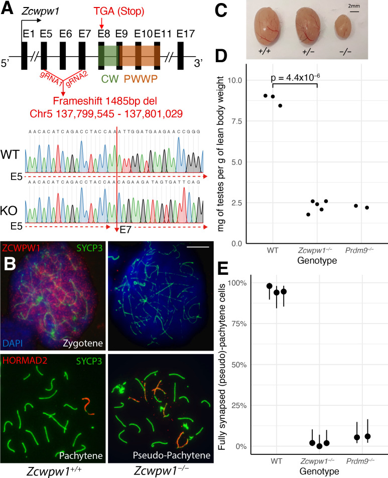
Figure 3—source data 1. Fertility measures in WT (+/+), Zcwpw1-/- and Prdm9-/- males.
Fertility was assessed in mice ranging from 8 to 12 weeks of age through measurement of paired testes weight and sperm count. Mouse ID is consistent across Figure 3—source data 2 and Figure 4—source data 1.
Figure 3—source data 2. Breeding performance of Zcwpw1-/- females.
All females were crossed with a WT male. N/A, not applicable; TLL, total litter loss; *assigned a value of 0 as the number of pups born in the total counts.

Figure 3.

Figure 3—figure supplement 1. Loss of ZCWPW1 expression in Zcwpw1−/− mouse testis.

Figure 3—figure supplement 1.

(A) Testis protein extracts from adult (10–12 weeks old) B6 wild-type (WT), Zcwpw1−/− and Prdm9−/− were immunoprecipitated with an anti-ZCWPW1 antibody (2.7mg/IP), followed by western blot detection with the same antibody. Detection of beta-actin from protein extracts (100µg) shows equal input for IP. (B) Testis chromosome spreads from 9- to 10-week-old Zcwpw1+/+ and Zcwpw1−/− mice were immunostained with antibodies against the synaptonemal complex protein SYCP3, and ZCWPW1, and counterstained with DAPI to visualise nuclei. Developmental stages are indicated at the top. The top row of panels shows merged signals, and the bottom rows individual signals. For ease of comparison, the boundaries of the mid-Leptotene cell in the Zcwpw1+/+ sample are marked by a rectangle. Red arrows point to ZCWPW1 foci at the ends of the synaptonemal complex (white arrows in the single-channel image). The yellow arrow points to the XY body. These images are representative of the results obtained for three mice per genotype. Scale bar: 10 μm.
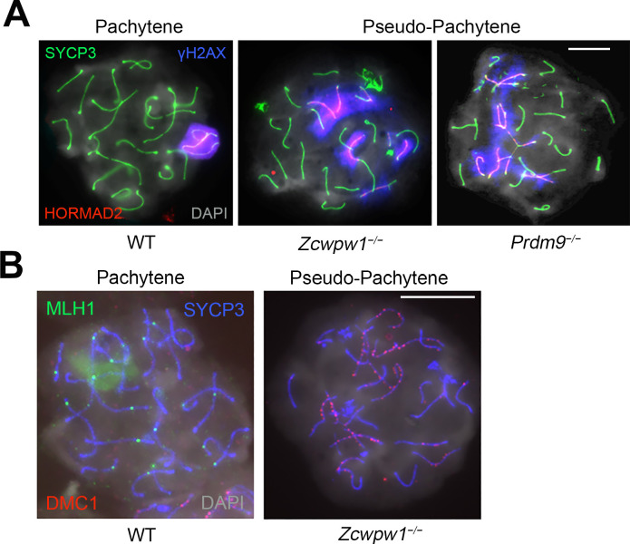
Figure 3—figure supplement 2. Asynapsis, and lack of XY body formation and crossover sites in Zcwpw1−/− mouse testis.

Figure 3—figure supplement 2.

Testis chromosome spreads from 9- to 12-week-old WT, Zcwpw1−/− and Prdm9−/− mice were immunostained with antibodies against SYCP3 (A–B), γ-H2AX (phosphorylated form) and HORMAD2 (A), or MLH1 and DMC1 (B), and counterstained with DAPI (A–B). WT Pachytene cells show full synapsis of all autosomes, an XY body strongly labeled by γ-H2AX and at least one (obligate) crossover site per chromosome labeled by MLH1. In contrast, many chromosomes are asynapsed and the XY body is absent in pseudopachytene cells from Zcwpw1−/− and Prdm9−/− mice; in the Zcwpw1−/− mutant, no MLH1 foci are observed either, and many DMC1 foci persist on asynapsed chromosomes. In the Prdm9−/− mutant, mispairing of homologues is evident by the formation of branched structures referred to as ‘tangled’ chromosomes in the text and Figure 3—figure supplement 2—source data 1. These images are representative of the results obtained for two (Prdm9−/−) to three mice (WT and Zcwpw1−/−) per genotype. Scale bar: 10 μm.
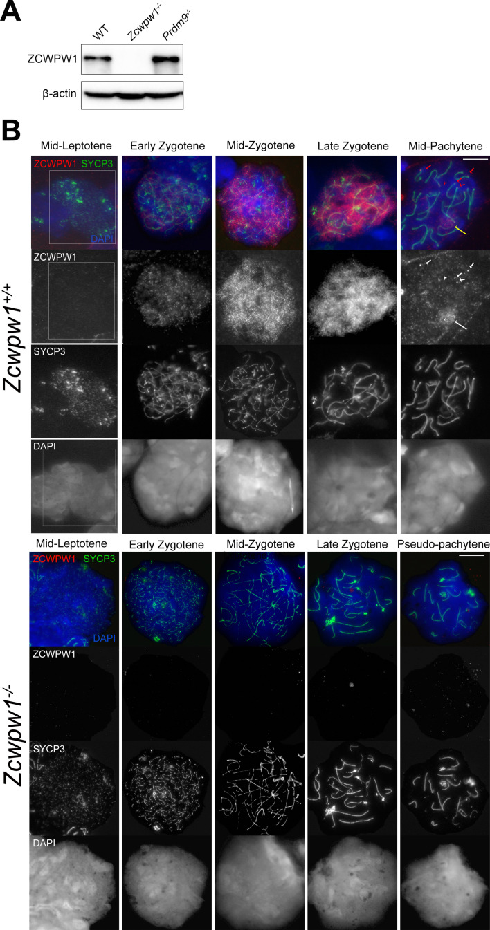
Figure 3—figure supplement 2—source data 1. Impaired synapsis in Zcwpw1-/- males.
The number of normal pachytene cells showing full synapsis of all autosomes and sex chromosomes (expressed as ‘% synapsis’ of all cells analysed) was determined by immunostaining of testis chromosome spreads against SYCP3, HORMAD2 and γ-H2AX (see images in Figure 3—figure supplement 2). The nature of the defects observed in cells with asynapsis was recorded as either ‘tangled’ (when chromosomes pair with the wrong partner, forming branched tangled structures); ‘multibodies’ strongly positive for HORMAD2 (when asynapsed chromosomes form multiple XY-like bodies which end up merging with each other, and with the XY body); or ‘split XY’ (when the X and Y sex chromosomes are found away from each other in different areas of the cell nucleus). Mouse ID is consistent across Figure 3—source data 1 and 2 and Figure 4—source data 1.