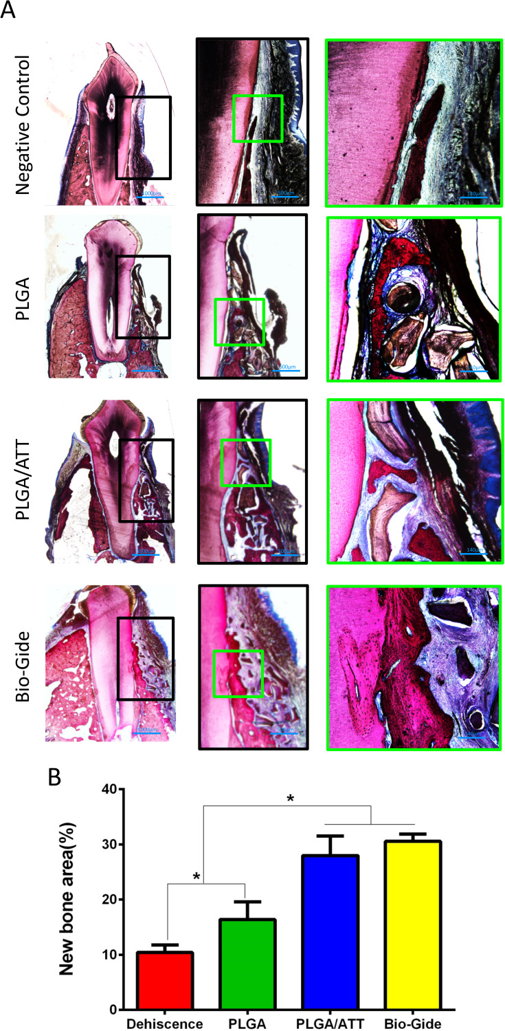
Figure 9.
Histological analysis of newly formed bone in buccal dehiscence defects 12 weeks post-implantation. (A) Non-decalcified specimens were sliced, and sections were stained with van Gieson’s picrofuchsin (Row 1, ×12.5; Row 2, ×40; Row 3, ×100). Red areas represent newly formed bone, blue areas indicate collagen fibers, and black areas represent undegraded remnants. (B) Percentage of new bone area (*p < 0.05).

