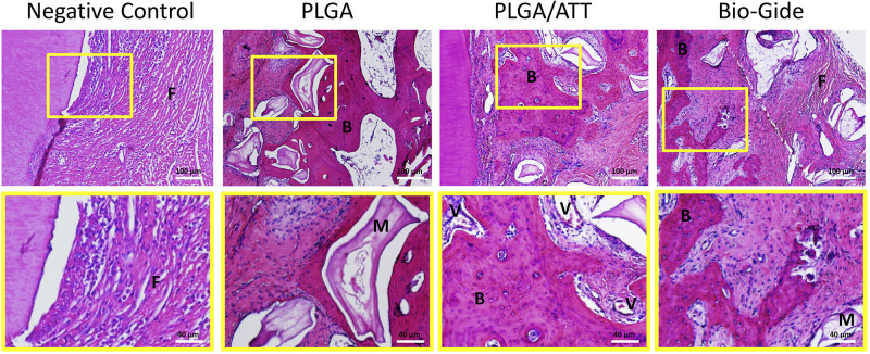
Figure 10.
Decalcified HE staining sections of the bone defect area in each group 12 weeks post-implantation. M indicates the residual materials; V, capillary vessels; B, thick bone trabeculae; M, residual materials; and F, fibrous tissue. Lower magnification images (×100) are shown in the upper panel. The region of interest is indicated by the yellow box and its magnified image (×250) is presented in the lower panel.
Abbreviations: HE, hematoxylin and eosin; PLGA, poly (lactic-co-glycolic acid) (PLGA); ATT, attapulgite.

