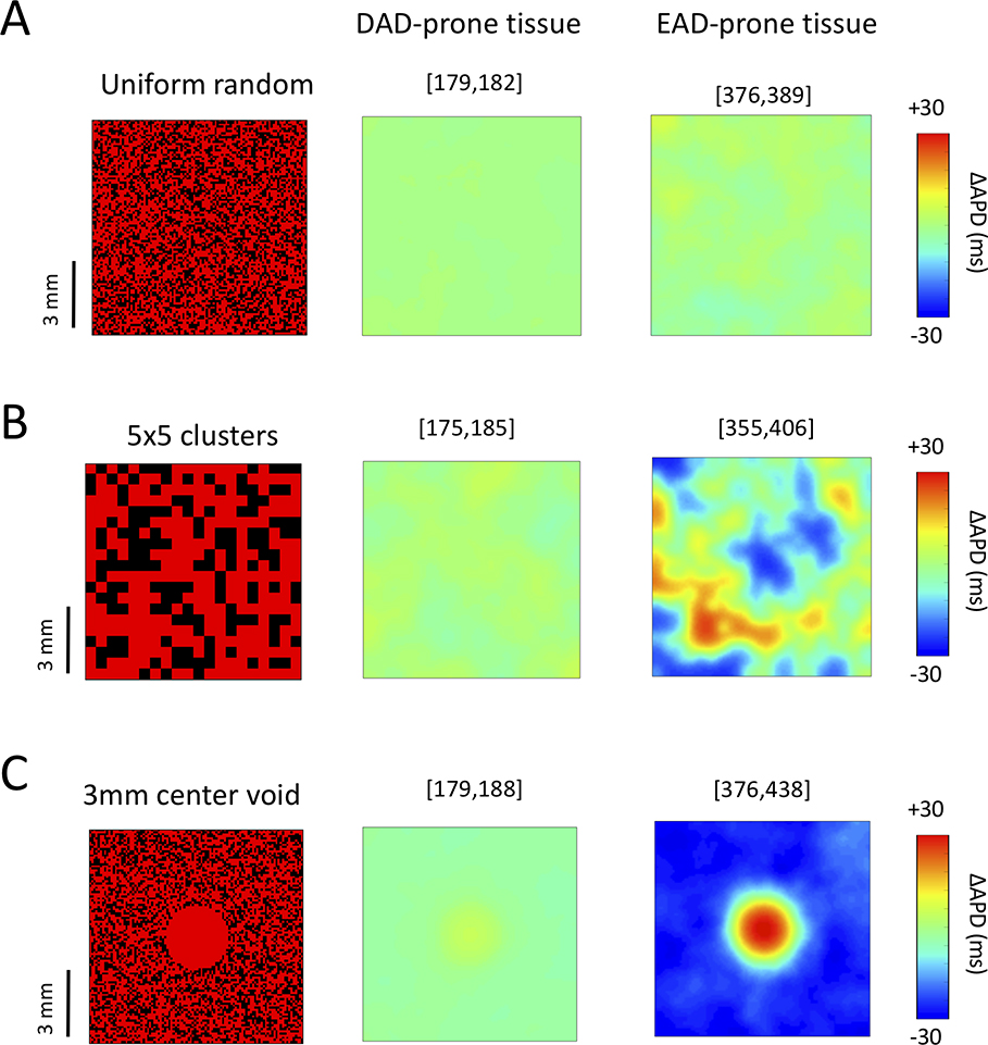
Figure 6.
Effect of GK1-enhanced stabilizer cells on APD dispersion. Stabilizer cells with GK1 enhanced 4-fold (40% of all cells) were randomly distributed in 2D tissue (100×100 cells) containing either DAD-generating cells (Gspon = 0.2 ms−1 and 50 ms standard deviation in DAD timing, middle panels) or EAD-generating cells (right panels). ΔAPD maps (difference from the mean tissue APD) are shown for: (A) Uniform random spatial distribution of single stabilizer cells; (B) Random distribution of 5×5 stabilizer cell clusters: (C) A 3 mm central defect devoid of stabilizer cells. Numbers in brackets above each panel indicate the tissue APD range in ms.

