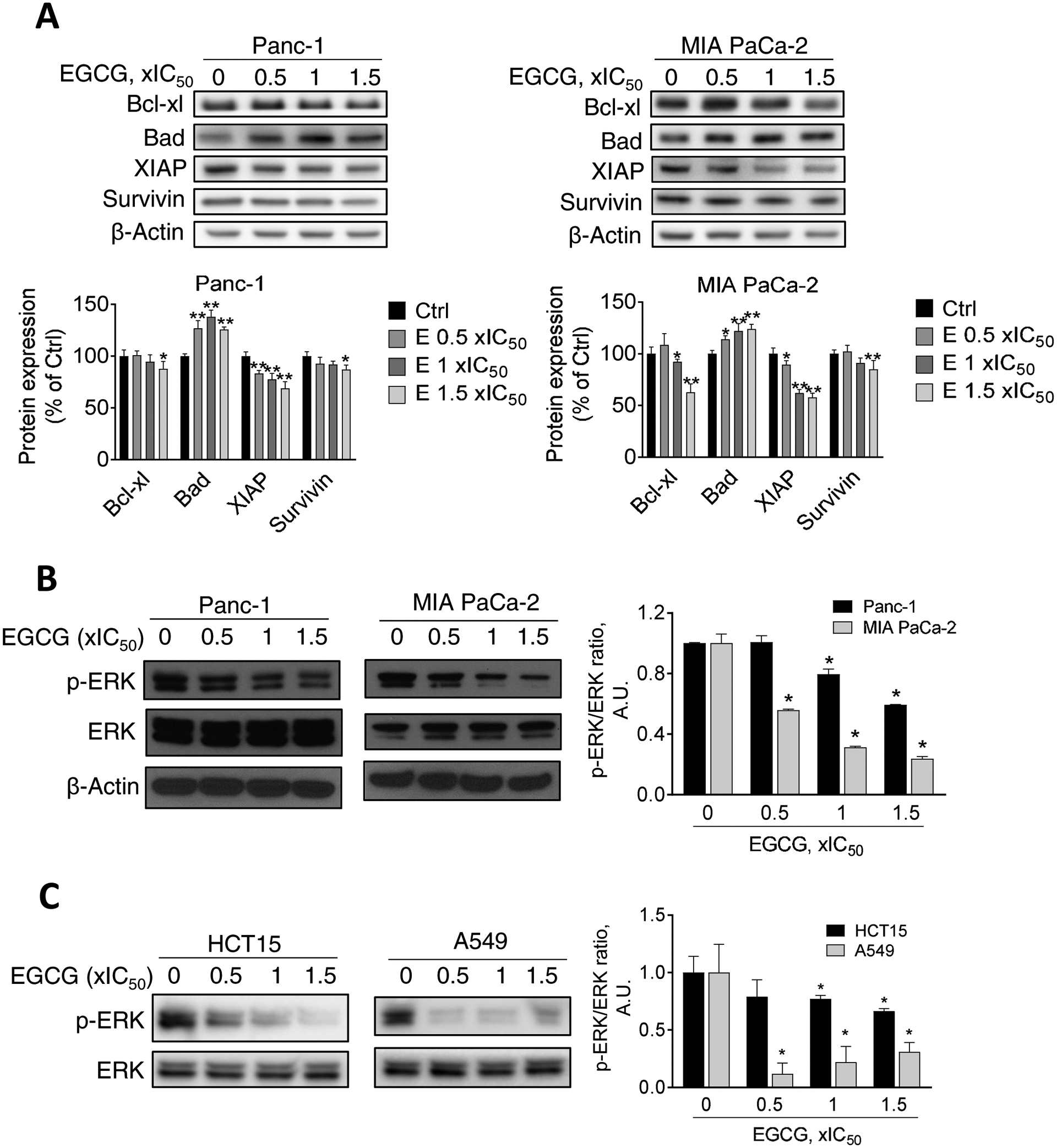
Figure 3. Effect of EGCG on apoptosis-related protein expression and ERK phosphorylation in multiple human cancer cells.

A: EGCG modulates the Bcl-2 family and XIAP family protein expression. Immunoblots for Bcl-xL, Bad, XIAP, and survivin in total cell protein extracts from Panc-1 and MIA PaCa-2 cells treated with escalating concentrations of EGCG, as indicated, for 24 h. Loading control: β-Actin. Bands were quantified and results are expressed as percentage of control. *p<0.05, **p<0.01 vs. control. B: EGCG reduces ERK phosphorylation in Panc-1 and MIA PaCa-2 cells, in a concentration-dependent manner. Results are expressed as the p-ERK/ERK ratio, normalized to control. The sample labeled as “0” refers to an untreated control. *p<0.05, vs. control. C: EGCG reduces ERK phosphorylation in HCT15 and A549 cells. Immunoblots for p-ERK, and ERK in total cell protein extracts from HCT15 and A549 cells treated with escalating concentrations of EGCG (0.5x, 1x and 1.5x IC50) for 24 h. Results are expressed as the p-ERK/ERK ratio, normalized to control. The sample labeled as “0” refers to an untreated control. *p<0.05, vs. control.
