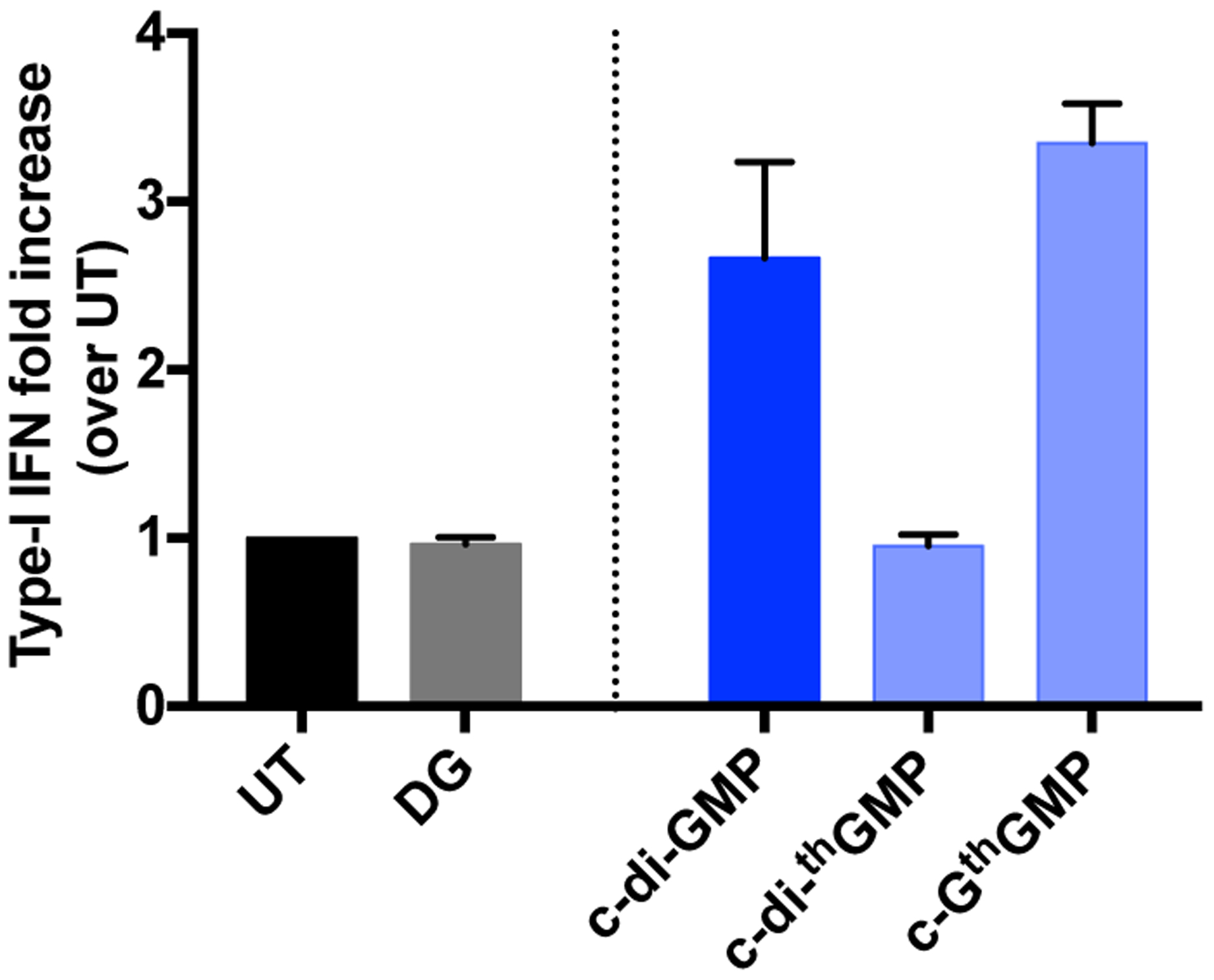
Figure 2.

Type-I IFN induced by CDNs in THP-1 cells. THP-1 cells were seeded at a density of 100,000 cells/well in a 96-well cell culture plate and differentiated with 25 nM of PMA for approximately 20 h prior to treatment with CDNs. Cells were transfected with 5 μM of CDNs in a permeabilization buffer containing 5 μg/mL of digitonin, then washed and incubated in RPMI medium with 2% FBS at 37°C for 4 h. 50 μL of cell culture supernatant per well was transferred to 150 μL of HEK-Blue IFN α/β reporter cells seeded at 50,000 cells/well in a 96-well cell culture plate and incubated at 37 overnight. The reporter cells were spin down the next day, and 50 μL of cell culture supernatant per well was transferred to a 96-well plate and added with 150 μL of QUANTI-Blue™ SEAP detection medium (InvivoGen). The samples were then incubated at 37°C for 1 h 20 min before absorption was measured at 640 nm. The absorption signal of each sample was normalized to untreated samples. Two independent assays were done in duplicates or triplicates. Error bars indicate SD.
