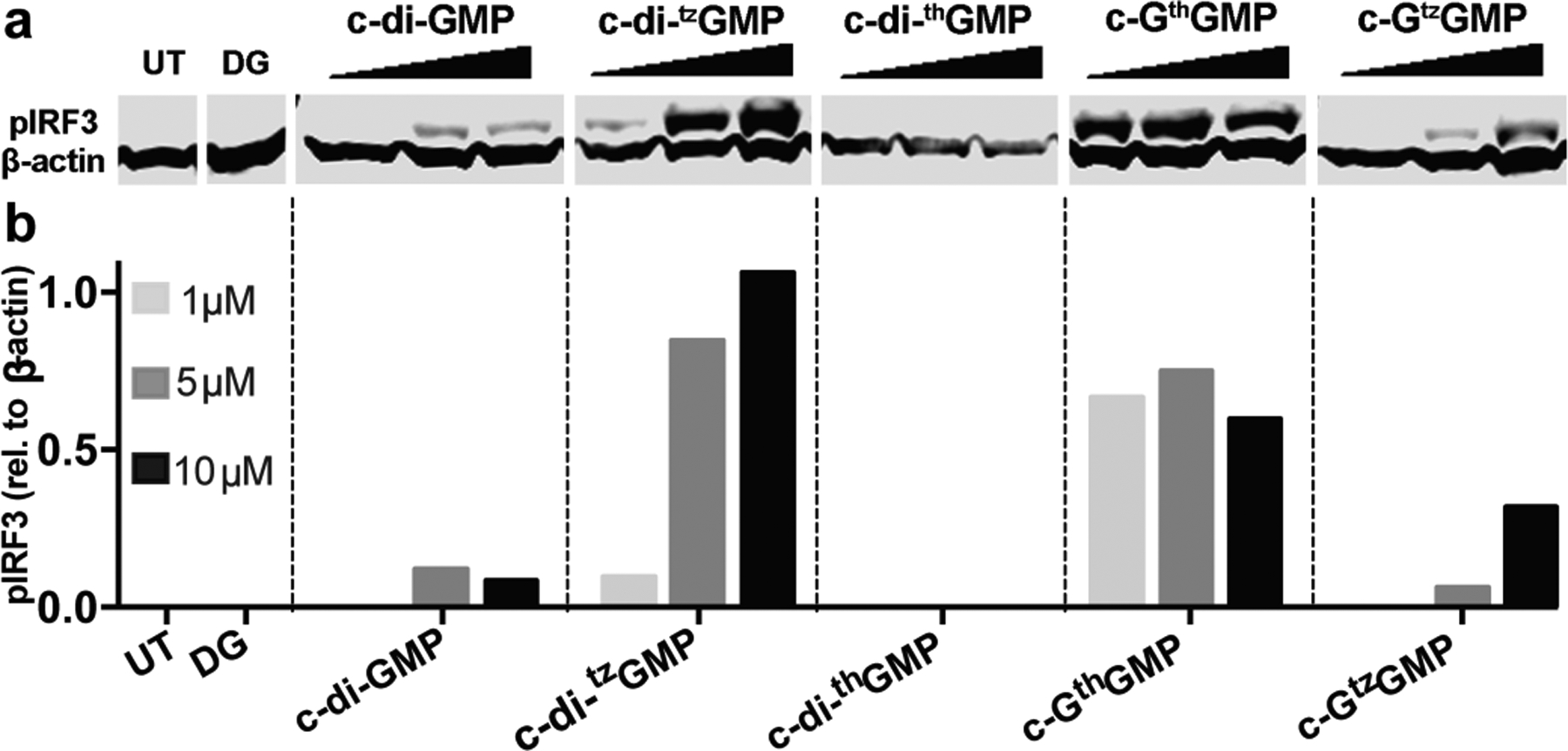
Figure 3.

IRF3 phosphorylation induced by c-di-GMP and its analogues. (a) IRF3 phosphorylation induced by c-di-GMP analogues. 1, 5 and 10 μM of each CDN was used to transfect RAW 264.7 cells. Cells were lysed with NP-40 lysis buffer 2 h post transfection, 20 μg of total protein was loaded on SDS-polyacrylamide gel. Proteins were transferred to PVDF membrane after gel electrophoresis, and immunoblotted against pIRF3 and β-actin. (b) quantification of western blot. Y-axis indicates relative intensity of pIRF3 compare to β-actin.
