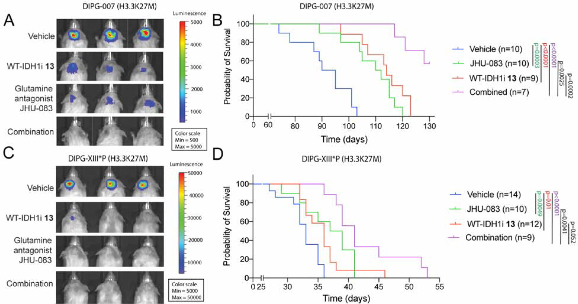
Figure 6. Inhibition of IDH1 and glutamine metabolism is therapeutic in vivo.

(A–B) Representative bioluminescence images (a) and Kaplan-Meier analysis (b) from animals implanted with H3.3K27M DIPG-007 cells in the pons and treated for four weeks (see Fig S6b) with vehicle (n=10), WT-IDH1i 13 (n=9) or the glutamine antagonist JHU-083 (n=10) or both (n=7).
(C–D) Representative bioluminescence images (c) and Kaplan-Meier analysis (d) from animals implanted with H3.3K27M DIPG-XIII*P cells in the pons and treated for three weeks (see Fig S6c) with vehicle (n=14), WT-IDH1i 13 (n=12) or the glutamine antagonist JHU-083 (n=10) or both (n=9).
See figure S6 for treatment details.
