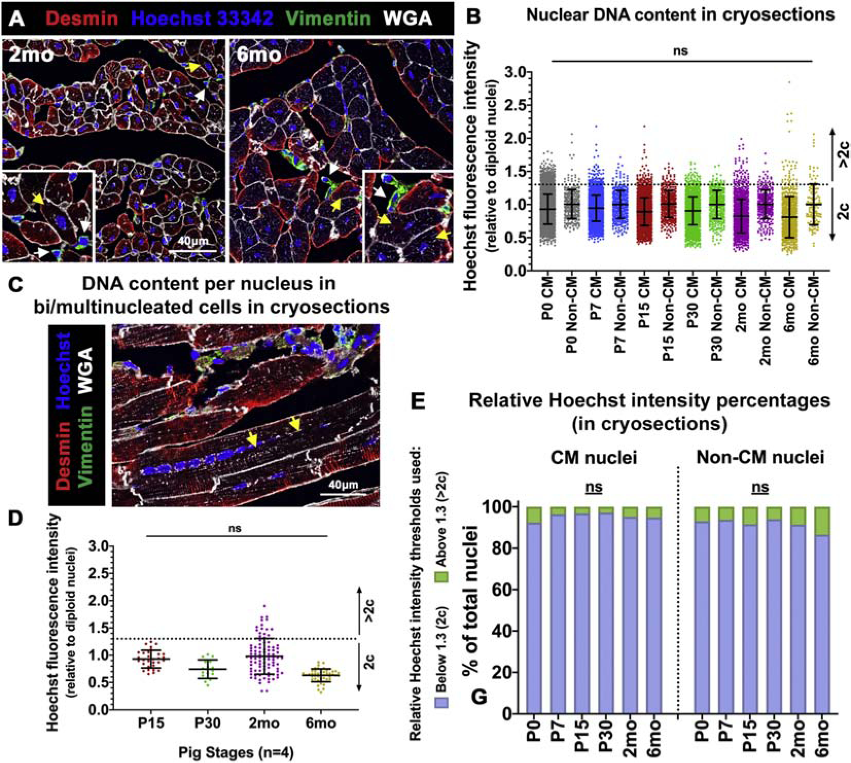
Figure 4. Pig CM nuclei remain predominantly diploid after birth.

(A) Representative images of ventricular tissue cryosections stained with Hoechst 33342 for CM nuclear intensity assessment (yellow arrows), with Vimentin-positive non-CM cell nuclei (white arrows) utilized as diploid (2c) control. (B) Hoechst fluorescence intensity per CM nucleus, relative to Vimentin-positive non-CM nuclear Hoechst intensities, was calculated per image. Thresholds for 2c and >2c were determined based on standard deviation of non-CM (2c) nuclei (>100 CMs analyzed per stage). (C) Representative image of a multinucleated CM (yellow arrows) in longitudinal tissue section stained with Desmin, Hoechst, and WGA, for nuclear intensity measurements. (D) Hoechst fluorescence intensity per nucleus in multinucleated CMs was measured at P15–6mo in cryosections. (E) Percentages of CM and non-CM nuclei with relative Hoechst intensities at 2c or >2c, as determined in tissue longitudinal cryosections. Data are mean ± SEM, with ns=not significant determined compared to P0 by Brown-Forsythe and Welch ANOVA tests, in n=4 pigs per stage.
