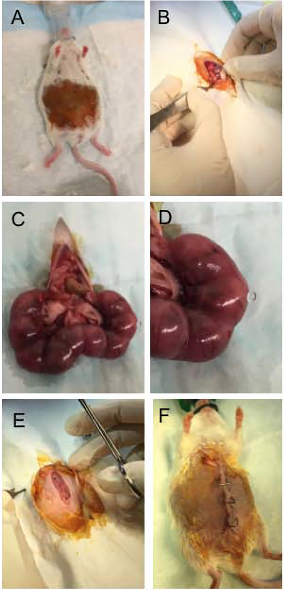
Figure 1: Drainage of amniotic fluid surgery.

Pregnant females at day 15.5 gestation underwent surgery under isoflurane anesthesia (A). The peritoneum was opened (B), and embryos were removed (C). One third of the total number of embryos underwent a puncture of the amniotic sac with a 25G needle (D). Remaining embryos were left as controls to prevent spontaneous abortion or preterm labor. After the perforation, embryos were returned to their initial position. The peritoneal cavity was flushed with saline and closed with sutures (E), the skin was closed with clips (F). Pregnant females were then sacrificed at E16.5, E17.5, and E18.5 (24, 48 and 72 hours post-surgery).
