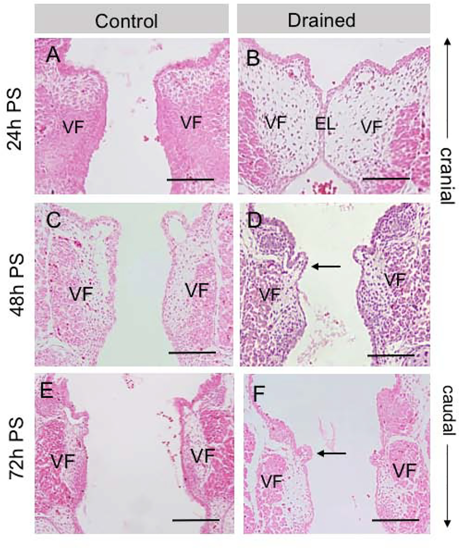
Figure 2: Characterization of VF morphology.

(A, B) Hematoxylin-eosin staining demonstrating a completely disintegrated EL in control embryos (A) and a visible EL in drained VF 24h PS (B). (C, D) Hematoxylin-eosin staining showing completely resorbed EL 48h PS in both control (C) and drained embryos (D). In drained VF, polyp-like structures appear in the mid-membranous region on the right and left VF (black arrow). These structures are lined with a very thin one-layer VF epithelium. (E, F). Hematoxylin-eosin staining demonstrating separated VF in control (E) and drained samples (F). Polyp-like structures persist in the mid-membranous VF region (black arrow). Scale bar = 100μm. Abbreviations: EL, epithelial lamina; h, hour; PS, post-surgery; VF, vocal fold.
