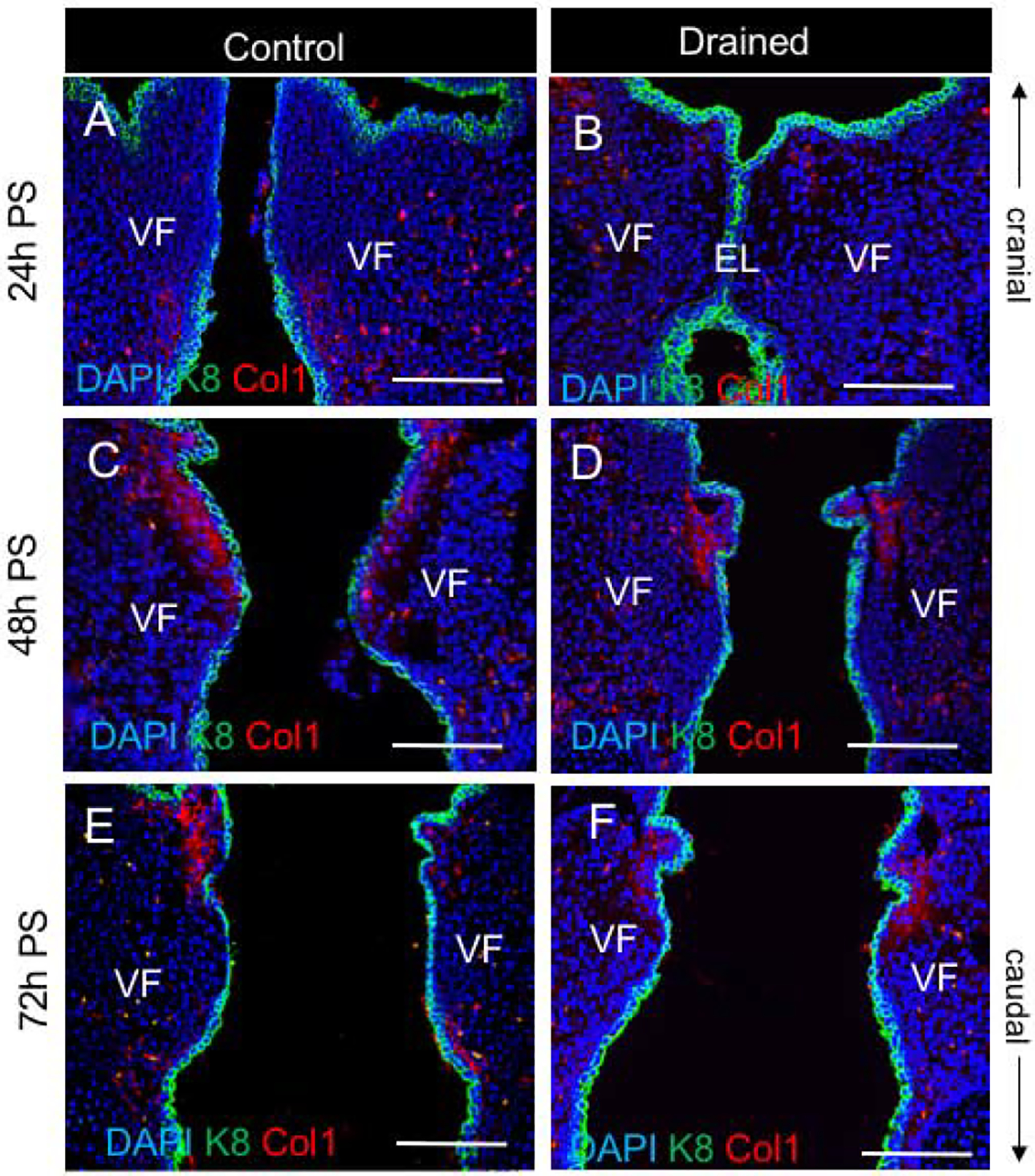
Figure 5: Deposition of Collagen1 in the lamina propria during the EL recanalization.

(A, B) Double staining for Col1 (red) and Cytokeratin K8 (green) in control (A) and drained embryos (B) 24h PS. In control VF, Col1 is detected particularly in the subglottic VF region. In drained VF, Col1 staining is reduced. (C - F) Double staining for Col1 (red) and Cytokeratin K8 (green) in control (C, E) and drained embryos (D, F) 48h and 72h PS, respectively. Collagen 1 expression was detected in both control and drained VF. Scale bar = 100μm. Abbreviations: EL, epithelial lamina; h, hour; PS, post-surgery; VF, vocal fold.
