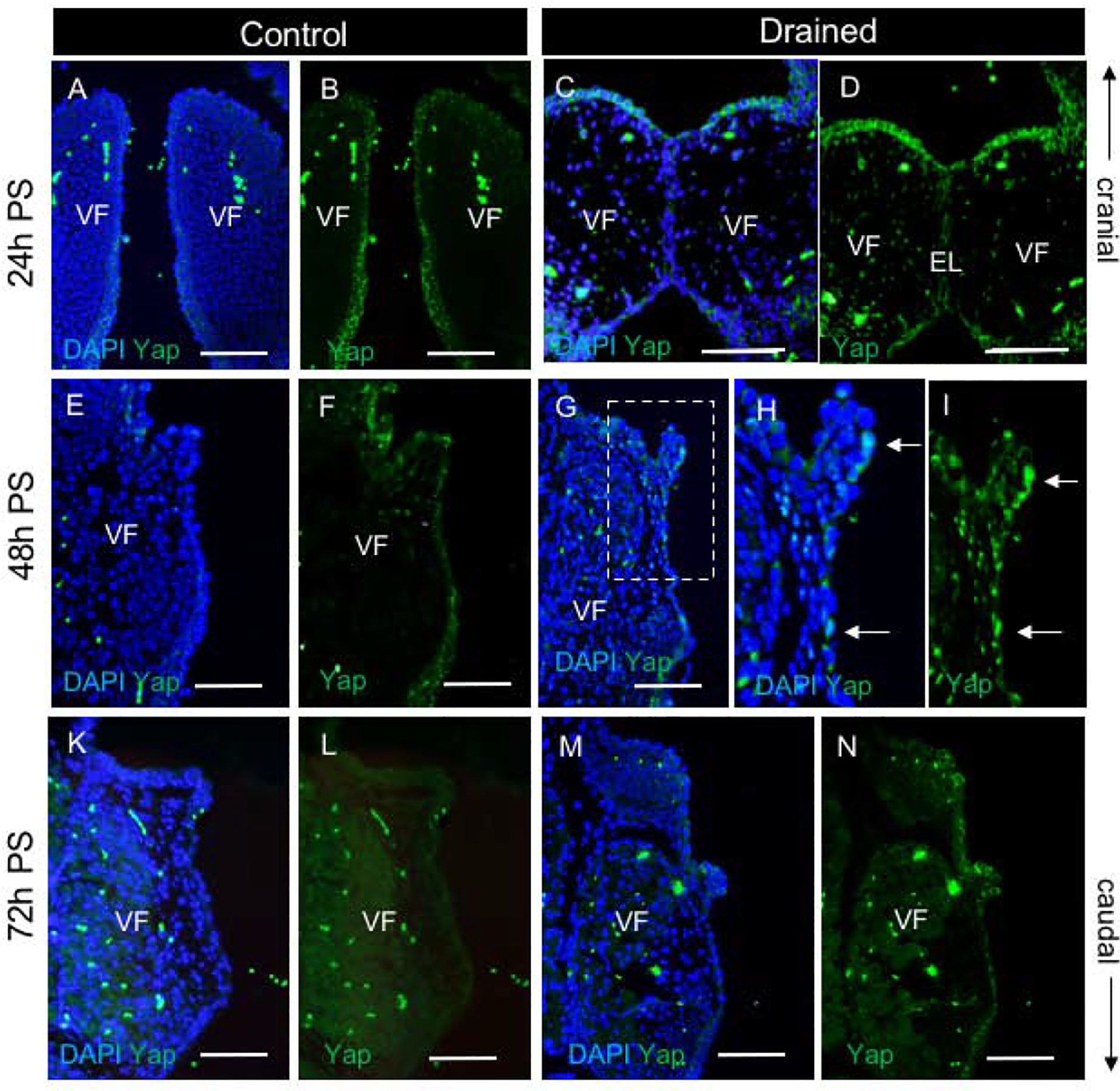
Figure 8: Yap expression during EL recanalization.

(A-D) Staining for Yap (green) in control (A, B) and drained embryos (C, D) showing cytoplasmic localization of Yap in VF epithelial cells. A single channel Yap expression in control (B) and drained embryos (D). (E-I) Staining for Yap (green) in control (E, F) and drained embryos (G-I) showing translocation of Yap into the nucleus in VF epithelium lining the polyp-like structure 48h PS (white solid arrows). A bracketed region in the panel of G is magnified in the panels of H and I. A single channel Yap expression in control (F) and drained embryos (I). (K-N) Staining for Yap (green) in control (K, L) and drained embryos (M, N) showing weak cytoplasmic Yap expression in the VF epithelium in control and drained vocal folds at 72h PS. A single channel Yap expression in control (L) and drained embryos (N). Scale bar = 100μm. Abbreviations: EL, epithelial lamina; h, hour; PS, post-surgery; VF, vocal fold.
