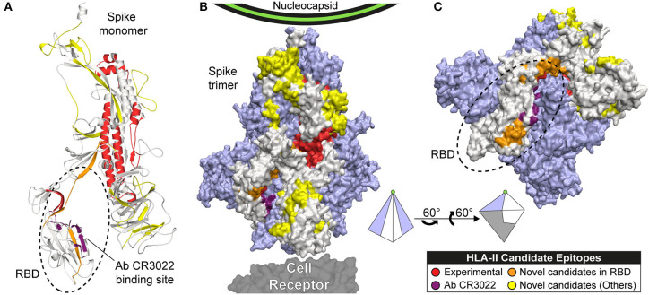
Figure 4.
Candidate epitopes in the 3D structure of the Spike (S) protein of SARS-CoV-2. This figure shows our HLA-II candidate epitopes with experimental evidence (red), without experimental evidence and CS ≥ 6 (orange: located in RBD, yellow: located in other regions), and the binding site of the Ab CR3022 (violet). Candidate epitopes were mapped to the Spike monomer (A), and the Spike trimer (B and C). The monomer is represented in beige, and the other 2 subunits in light purple. (C) Rotation of the trimer showing the RBD region. A pyramid representing the trimer is shown as visual aid to represent the rotation.

