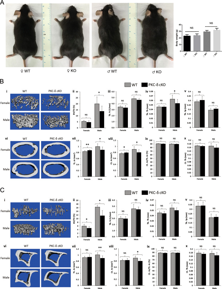
Fig. 1. Micro-CT analysis of hind limbs revealing an osteoporotic phenotype in 12-week-old mice with PKC-δ conditional knockout in B cells.
a Representative images and body weight of the mice at 12 weeks of age (WT wild-type, KO knockout). NS = non-significant compared with WT controls; b, c Representative 3D reconstructions of trabecular and cortical bone and bone parameters assessed by micro-CT in distal femur (b i–x) and proximal tibia (c i–x) in age- and sex-matched WT and PKC-δ conditional knockout (cKO) mice, respectively (male WT n = 6, male cKO n = 7, female WT n = 7, female cKO n = 7). Trabecular bone parameters (b ii–v and c ii–v) are shown as trabecular bone volume fraction (BV/TV, %; b ii and c ii), trabecular number (Tb.N, 1/mm; b iii and c iii), trabecular thickness (Tb.Th, mm; b iv and c iv) and trabecular separation (Tb.Sp, mm; b v and c v). Micro-CT analysis of cortical bone parameters (b vii–x and c vii–x) are shown as total cortical area (Tt.Ar, mm2; b vii and c vii), cortical bone area (Ct.Ar, mm2; b viii and c viii), cortical area fraction (Ct.Ar/Tt.Ar, %; b ix and c ix) and cortical thickness (Ct.Th, μm; b x and c x). Data are presented as mean ± SD. *p < 0.05, **p < 0.01, NS = non-significant compared with WT controls.

