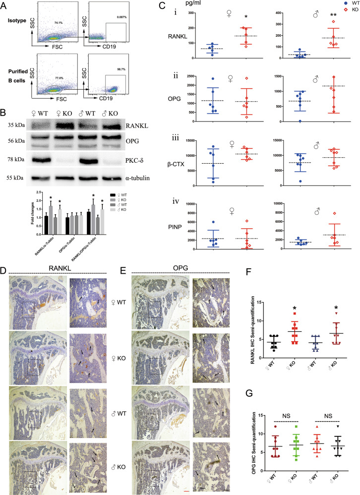
Fig. 4. PKC-δ deficiency in B cells elevated RANKL/OPG ratio in both cell culture and serum and increased RANKL expression in the trabecular bone.
a Flow-cytometry plots representing the purity of B cells after sorting; b Expression of PKC-δ, OPG, and RANKL in B cells from WT and PKC-δ cKO mice was detected by western blotting, with semi-quantitative analysis; c Blood was obtained from the fundus vein of WT and PKC-δ cKO mice. The concentrations of RANKL, OPG, β-CTX, and PINP were determined by ELISA in the serum. Each data point was from an individual mouse and means were indicated by dashed horizontal lines. d, e Representative images of RANKL (d) and OPG (e) immunohistochemistry stained tibia sections of 3-month-old PKC-δ cKO and age-sex-matched wild-type mice. Higher magnification micrograph of area within square in d, e is presented at the right side of each. Arrows indicate the positive staining of osteoclasts in the trabecular bone within the tibia. Red bar and black bar represent 200 μm and 50 μm, respectively. f, g Semi-quantitative analysis of RANKL (f) and OPG (g) immunohistochemistry staining. Bar charts represent mean ± SD. *p < 0.05, **p < 0.01, NS = non-significant compared with WT control group.

