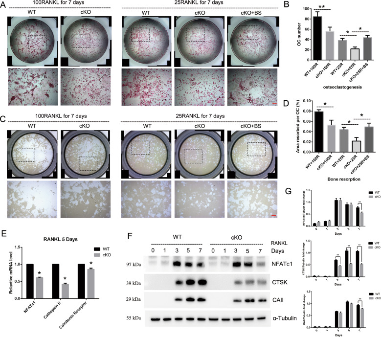
Fig. 5. Deletion of PKC-δ selectively in B cells resulted in suppressing osteoclast differentiation and function, but B-cell supernatant co-culture exerted reversal effect on osteoclast formation and activation.
a Representative images of osteoclasts with TRAP staining after 100 ng/ml or 25 ng/ml RANKL (with and without BS co-culture) induction for 7 days, the square in the upper images of each well indicate where the lower images were captured. Bar represents 200 μm; b Quantification of the number of osteoclasts, TRAP-positive cells containing three or more nuclei were counted as osteoclasts; c Representative images of eroded areas in hydroxyapatite-coated plates after 100 ng/ml or 25 ng/ml (with and without BS co-culture) RANKL stimulation for 5 days, the square in the upper images of each well indicate where the lower images were captured. Bar represents 200 μm; d Quantitative analysis of the resorbed proportion per osteoclast by measuring the area of the mineral coating removal; e–g Gene transcription (e) and protein expression f–g analysis of osteoclast-specific markers NFATc1, Cathepsin K (CTSK), Calcitonin Receptor and Carbonic Anhydrase II (CAII) by RT-PCR and western blotting. BS B-cell supernatant. All the experiments were carried out in triplicate from 12-week-old male WT and cKO mice, results are presented as mean ± SD. *p < 0.05, **p < 0.01 vs. WT control group.

