
Figure 2.
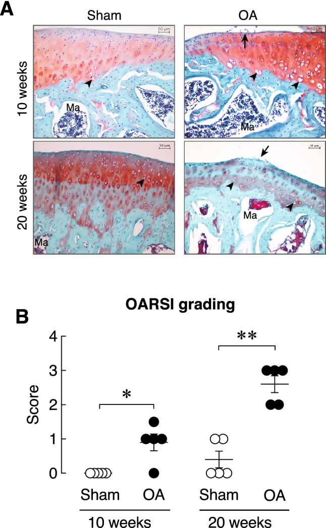
(A) Representative photomicrographs of right knee joint obtained from sham and OA rats at 10 and 20 weeks after OA induction stained with Safranin O/Fast green staining. Red color indicates articular cartilage. Green color indicates subchondral bone. Arrow indicates mild abrasion on the articular cartilage surface of tibial plateau. Arrow head indicates articular chondrocytes. Ma, marrow. Scale bars are 50 µm. (B) OARSI average grading score in the right knee joint obtained from sham and OA rats at 10 and 20 weeks after OA induction (n = 5 per group). *P < 0.05, **P < 0.01 vs. corresponding Sham group.