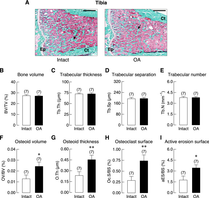
Figure 4.
(A) Representative photomicrographs of Goldner’s trichrome stained tibial growth plate obtained from 20-week OA rat and its intact tibiae. Epiphyseal plate (Ep), and cortical shell (Ct) were identified. Mineralized trabeculae (arrows) and marrow cells were stained green and red, respectively. Bars, 1 mm. Microstructural analysis of tibial metaphysis in OA rats and its intact leg at 20 weeks after OA induction as determined by bone histomorphometry. (B) trabecular bone volume normalized by tissue volume (BV/TV), (C) trabecular thickness (Tb.Th), (D) trabecular separation (Tb.Sp), (E) trabecular number (Tb.N), (F) osteoid volume (OV) normalized by bone volume (BV), (G) osteoid thickness (O.Th), (H) osteoclast surface (Oc.S) normalized by bone surface (BS), and (I) active erosion surface (aES) normalized by BS. Numbers of animals in each group are shown in parentheses. *P < 0.05 and **P < 0.01 vs. intact leg.

