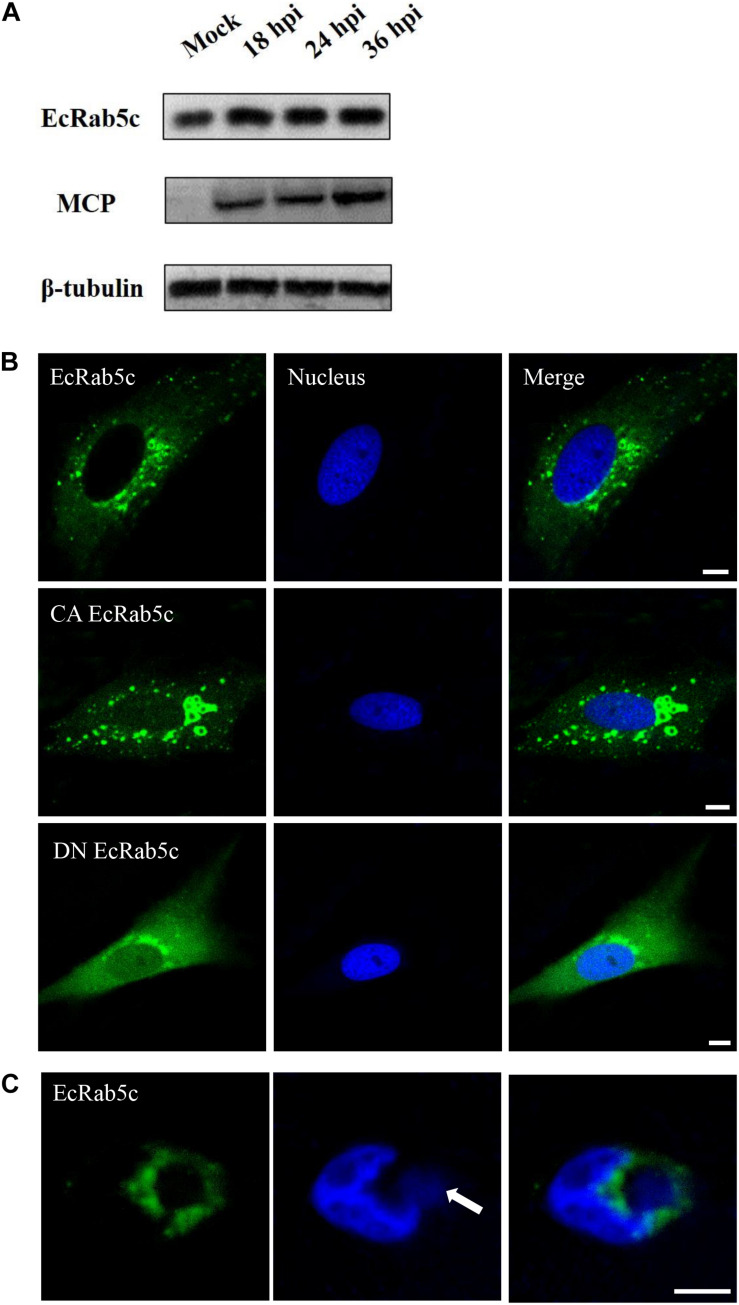
FIGURE 1.
Expression patterns and subcellular distribution of EcRab5c. (A) EcRab5c and viral protein level were detected at different times post SGIV infection. GS cells were collected at 18, 24, and 36 hpi for Western blotting, and β-tubulin was used as the internal control. (B) Distribution pattern of EcRab5c, CA EcRab5c, and DN EcRab5c. GS cells were transfected with pEGFP-EcRab5c, pEGFP-CA EcRab5c, and pEGFP-DN EcRab5c, respectively. Scale bars are 5 μm. (C) EcRab5c located near the virus factory. GS cells transfected with pEGFP-EcRab5c were infected with SGIV and fixed at 24 h. The nucleus and virus factory were stained by Hoechst 33342. The white arrow shows the virus factory. Scale bars represent 10 μm.

