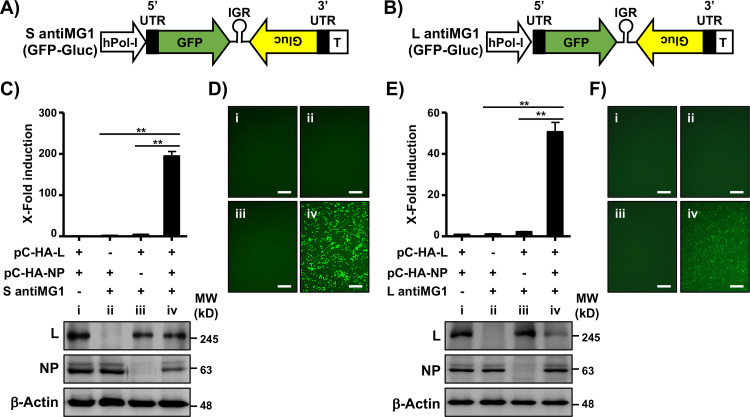
FIG 2.
Verification of TCRV BEI TRVL-11573 S and L NCRs using an MG assay. (A and B) Schematic representation of TCRV S (A) and L (B) MG reporter-expressing plasmids. The backbone of the TCRV TRVL-11573 S and L, including 5′ and 3′ NCRs (black boxes) and IGRs (loops), was used to generate the MG reporter-expressing plasmids under the control of an hPol-I promoter and a mouse Pol-I (mPol-I) terminator I (T) sequence in antigenome (antiMG1) polarity. GFP was used to replace the viral NP (S segment) (A) and L (L segment) (B). Gluc was used instead of the GPC (S segment) (A) and Z (L segment) (B) viral genes. (C and D) TCRV S segment antiMG1 activity. HEK293T cells (6-well format, 106 cells/well, triplicates) were cotransfected with the indicated plasmids, together with pSV40-Cluc to normalize transfection efficiencies, and Gluc and Cluc activities in the tissue culture supernatants (C, top), and GFP expression (D) were determined at 48 h posttransfection. Lanes i to iv correspond to the plasmid combinations shown in panel C for HEK293T cell transfection. Bars, 100 μm. The changes in fold induction were calculated by normalizing the ratio of Gluc/Cluc in cells transfected in the absence of pCAGGS-HA-L to 1. Expression of TCRV L and NP from pCAGGS plasmids in transfected cells was analyzed by Western blotting using a monoclonal antibody against the HA epitope tag. A monoclonal antibody against β-actin was used as a loading control. Molecular weight (MW) markers in kilodaltons (kD) are indicated on the right (C, bottom). (E and F) TCRV L segment antiMG1 activity. HEK293T cells (6-well format, 106 cells/well, triplicates) were cotransfected with the indicated plasmids, together with pSV40 Cluc to normalize transfection efficiencies, and Gluc and Cluc in tissue culture supernatants (E, top) and GFP expression (F) were determined at 48 h posttransfection. Lanes i to iv correspond to the plasmid combinations shown in panel E for HEK293T cell transfection. Bars, 100 μm. The changes in fold induction were calculated by normalizing the ratio of Gluc/Cluc in cells transfected in the absence of pCAGGS-HA-L to 1. Expression levels of TCRV L and NP in transfected cells were analyzed by Western blotting using a monoclonal antibody against the HA epitope tag. β-Actin was used as a loading control. MW are indicated on the right (E, bottom). Data are means and SD. **, P < 0.01.

