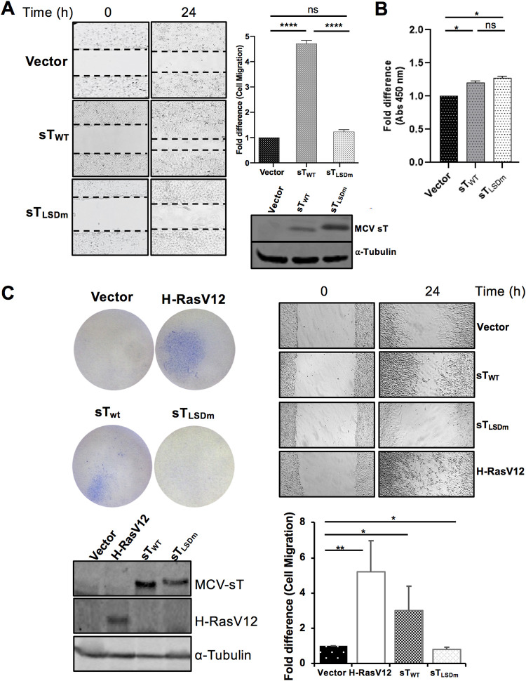
FIG 3.
MCV sT induces cell motility in an LSD-dependent manner. (A) MCV sT-induced cell migration is LSD dependent. Scratch assay. Poly-l-lysine-coated 6-well plates were seeded with U2OS cells and transfected with either a vector control or sTWT or sTLSDm plasmids. Migration of cells toward the scratch was observed over a 24-h period, and images were taken using a Revolve 4 fluorescence microscope (Echo Laboratories). Scratch assays were performed in triplicate and measured using Fiji ImageJ analysis software. Differences between means (P value) were analyzed using a t test with GraphPad Prism software. Protein expression was detected by immunoblot analysis to validate successful transfection using 2T2 antibody for sT antigen and α-tubulin, respectively. (B) No significant differences in cell proliferation were observed between cells expressing MCV sT within 24 h, indicating that cell proliferation does not interfere with the measurement of sT-induced cell migration. (C) MCV sT promotes rodent fibroblast cell migration. NIH 3T3 cells stably expressing an empty vector, H-RasV12, MCV sTWT, and sTLSDm were trypsinized and 2 × 10⁵ cells were used for transwell migration and scratch assay. H-RasV12 was used as a positive control. The experiments were performed two times, and the results were reproducible. The graph indicates the fold difference of migrated cells relative to the vector control sample. Protein expression was determined by immunoblotting.

