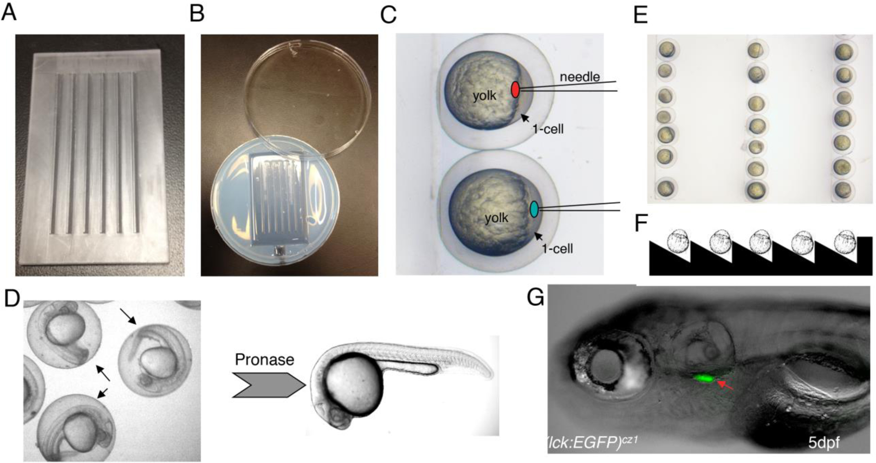
Fig.1. Microinjection and dechorionation for zebrafish embryos.

(A) Injection mold. (B) Injection chamber/plate. (C) Schematic of morpholino or plasmid injection. For morpholino knockdown, we inject the liquid (red spot) to the interface between cell and yolk. For plasmid overexpression, we directly inject the solution to the cytoplasm (green spot) at 1-cell stage. (D) Dechorionation process. The arrows indicate the chorions around the 24hpf embryos. (E) Alignment of the 1- cell stage embryos in the trenches of the injection plate. (F) Schematic of the vertical section of the injection plate. (G) Lateral view of a 5dpf Tg(lck:EGFP)cz1 embryo. Arrow indicates the EGFP+ T cells in the thymus.
