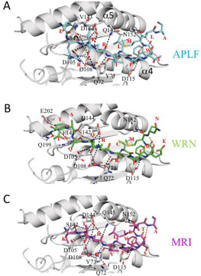
Figure 2. Detailed views of the KBM-Ku80 vWA structures showing H-bonds.
Hydrogen bonding between xlKu80 vWA (gray) and (A) APLF (cyan), (B) WRN (green), and (C) MRI KBM peptide (magenta) is indicated by black dotted lines. Interacting vWA residues are labeled, and each peptide residue type is indicated in red. In some cases, disordered sidechains have been truncated. Interpeptide H-bonds are shown as red dotted lines, and H-bonds mediated by water molecules are in blue.

