Image 2.
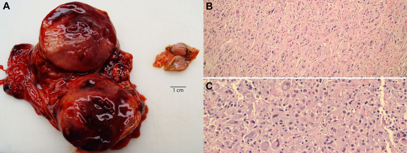
Adrenal tumors with bilateral pheochromocytoma. A, Photomicrograph of the enlarged, hemorrhagic adrenal mass (left) and a smaller adrenal nodule (right). Note the bright yellow rim of adrenal cortex around both tumors. B, Histologic sections from the right adrenal tumor show a neoplastic cell proliferation in Zellballen pattern. C, Sections of the larger left adrenal tumor show sheet-like proliferation of neoplastic cells. These cells demonstrate oval, hyperchromatic nuclei, mild to moderate nuclear pleomorphism, and abundant amphophilic granular cytoplasm. Hematoxylin and eosin, ×100.

