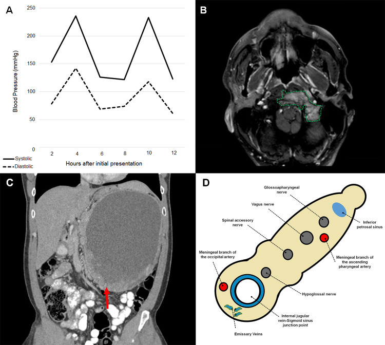
Figure 1.
A, Line plots of blood pressure fluctuations during the first 12 hours after initial presentation. B, Axial T1-weighted magnetic resonance image of the neck with contrast demonstrating avidly enhancing mass (outlined) within the left jugular foramen, extending medially and eroding the left aspect of the clivus. C, Coronal computed tomography of the abdomen showing large retroperitoneal soft tissue mass (arrow), likely the primary tumor, displacing loops of bowel inferiorly and medially. D, Cross-sectional schematic representation of structures passing through the jugular foramen.

