FIGURE 1.

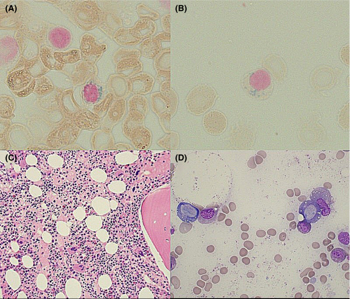
Bone marrow biopsy histopathology findings showing (A) ring sideroblast, (B) iron granules in plasma cell, (C) core biopsy with hypercellular marrow, and (D) vacuoles in erythroid and myeloid precursors

Bone marrow biopsy histopathology findings showing (A) ring sideroblast, (B) iron granules in plasma cell, (C) core biopsy with hypercellular marrow, and (D) vacuoles in erythroid and myeloid precursors