FIGURE 3.

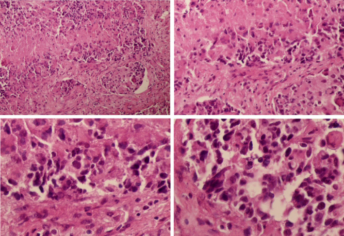
Pathological microscopic images of the lymph node biopsy showing: invasive proliferating anaplastic cells, large hyperchromatic nuclei and occasional bizarre mitotic figures, with no identified residual lymphoid tissue

Pathological microscopic images of the lymph node biopsy showing: invasive proliferating anaplastic cells, large hyperchromatic nuclei and occasional bizarre mitotic figures, with no identified residual lymphoid tissue