Figure 4.

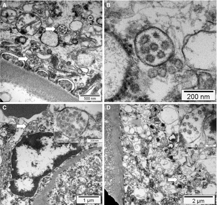
Electron microscopy findings. A,B, Podocyte cytoplasm with its foot processes on top of a glomerular basement membrane containing mitochondria (upper left corner) and multiple vesicles, two of which contain several small possible virus‐like particles with sizes between 70 nm and 110 nm (arrow). At higher magnification, the vesicles contain double membranes and the virus‐like particles show a ring of electron‐dense granules and a ragged outer contour (electron microscopy). C, An activated glomerular endothelial cell, and a vesicle close to the luminal border with virus‐like particles (arrow and insert), adjacent to an erythrocyte (electron‐dense structure at the top left) (electron microscopy). D, Cytoplasm of a proximal tubular epithelial cell on top of a basement membrane and adjacent collagen fibres (left side). The cytoplasm contains mitochondria, rough endoplasmic reticulum, and multiple vesicles, one of which contains virus‐like particles (arrow and insert, electron microscopy).
