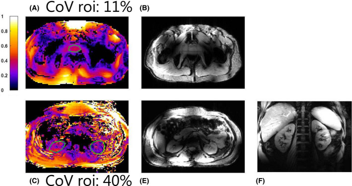
FIGURE 9.

In vivo image of the pelvis (top row) and abdomen (bottom row). B1‐maps (A and C) were acquired for both anatomies using the actual flip‐angle imaging method, as well as scout images (B and D). For the kidneys, a fat suppressed coronal image is shown (E)
