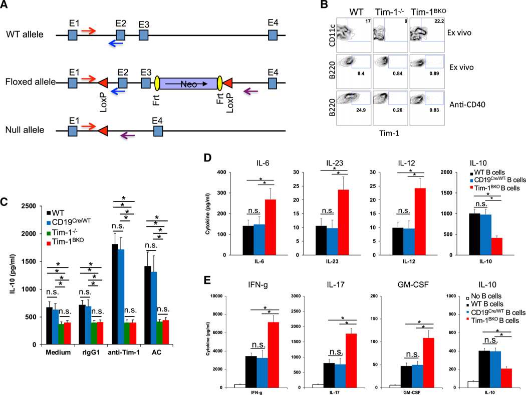
Figure 1. Generation of Tim-1BKO Mice.
(A) Strategy for generating Tim-1 floxed mice.
(B) Representative fluorescence-activated cell sorting (FACS) plots showing Tim-1 expression in dendritic cells (DCs) and B cells in spleens of 6- to 8-week-old mice (n = 6–8) ex vivo or in isolated B cells activated with anti-CD40 for two days.
(C) B cells isolated from 6- to 8-week-old mice (n = 5–6 per group) were cultured with anti-Tim-1, apoptotic cells (ACs), or controls. After 60 h, IL-10 production in culture supernatants was measured by ELISA.
(D) B cells isolated from 6- to 8-week-old mice (n = 5–8 per group) were cultured with lipopolysaccharide (LPS) for 40 h and then examined for their cytokine production in culture supernatants by BioLegend LEGENDplex.
(E) Foxp3− T cells from 6- to 8-week-old Foxp3-GFP knockin (KI) mice were cultured with B cells from WT or Tim-1BKO mice (n = 5 per group) plus soluble anti-CD3 for three days. Then, isolated T cells from the cultures were re-activated with plate-bound anti-CD3, and cytokine production in 40-h cultures was measured by BioLegend LEGENDplex. *p < 0.01; n.s., not significant. Data are represented as mean ± SEM.
See also Figure S1.

