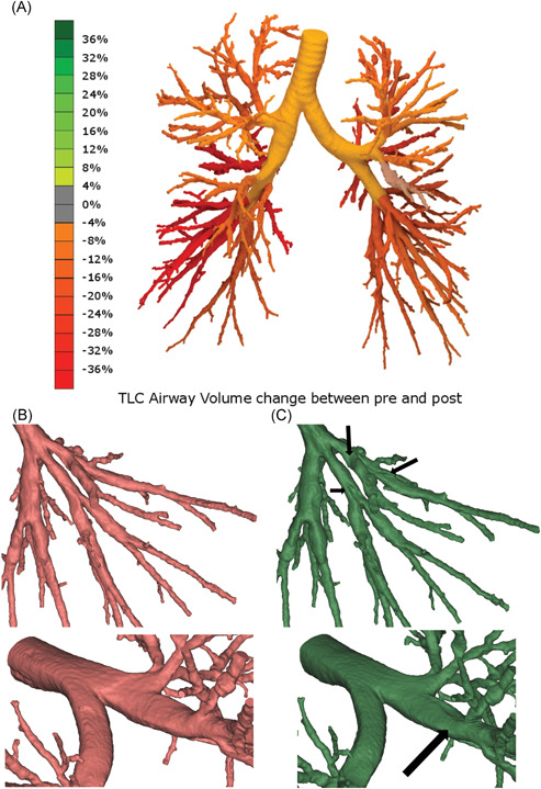
Figure 3.

A, Volume change in patient 009 airways: green color indicates dilation whereas red color indicates narrowing (due to mucus transportation). B, Detail of patient 009 distal (lower left lobe) airways before (red) and after (green) mHFCWO treatment; the arrows indicate mucus shift. C, Detail of patient 009 central airways before (red) and after (green) mHFCWO treatment; the arrow indicates mucus shift. HFCWO, high‐frequency chest wall oscillation [Color figure can be viewed at wileyonlinelibrary.com]
