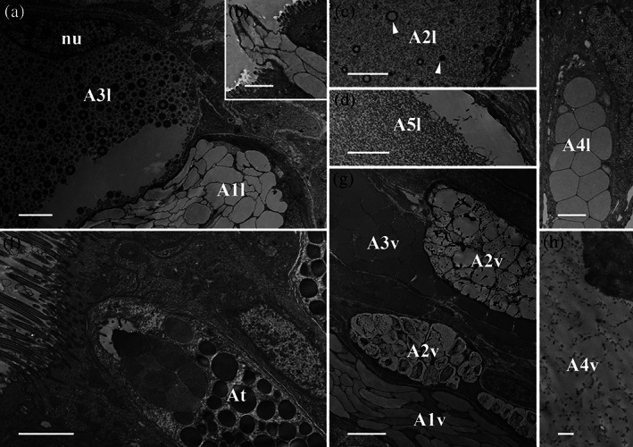
FIGURE 3.

Arion vulgaris, transmission electron micrographs of the lateral and ventral gland types. (a) The secretory cells of A1l and A3l could be clearly differentiated in an electron microscope; A1l bears electron‐translucent “ice floes” while the granules of A3l are spherical with several concentric rings and an electron‐translucent centre. The nucleus (nu) could be seen close to the secretory content of A3l. (b) The secretory content of A1l is extruded as a whole package. (c) The content of A2l consists of granulated material of different sizes (white arrowheads) and (d) that of A5l is homogeneous granular material. (e) Gland type A4l contains polygonal granules of homogenous material. (f) Gland type At, which occurs in the transition zone between the lateral and ventral sides, contains roundish granules merging near the apical pole. (g) In the ventral region, three different gland types (A1v, A2v, and A3v) can be observed, all appearing in high abundance and containing granules, different in size and appearance. (h) The secretory content of gland type A4v is finely grained with dark grained inclusions. Scale bars in a, c, d, f, and g = 2.5 μm, in image b and e = 2 μm and in image h = 0.5 μm
