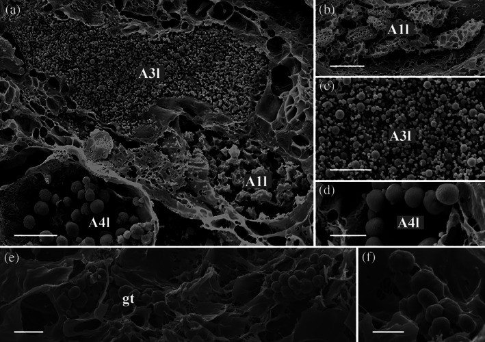
FIGURE 4.

Arion vulgaris, scanning electron micrographs of the lateral gland types. In the freeze‐dried tissue samples, (a) three gland types could be observed, which correlate in view of its granular content with the gland types A1l, A3l, and A4l (see Figure 3 for comparison). (b) The granules of gland type A1l are oval and appear sponge‐like. (c) Gland type A3l contains granules of different sizes, while (d) gland type A4l contains roundish, evenly sized granules. Scale bar in image a = 10 μm, in image b to c = 5 μm
