
FIGURE 2.
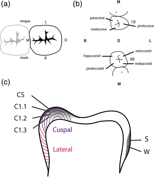
Sample locations in third molars. (a) Occlusal view (from above) indicating L = lingual (toward the tongue), M = mesial (toward the midline point/incisors of the dental arch), D = distal (opposite of mesial, toward the back of the dental arch), B = buccal (toward the cheek). (b) Cusp names (see also Table 1) for maxillary (above) and mandibular (below) third molars. (c) Profile view indicating CS = cusp surface, C1.1 = cusp layer 1, C1.2 = cusp layer 2, C1.3 = cusp layer 3, S = wall surface, and W = wall. Incremental enamel formation is indicated in purple (cuspal) and pink (lateral) lines (after Dean, 2000)