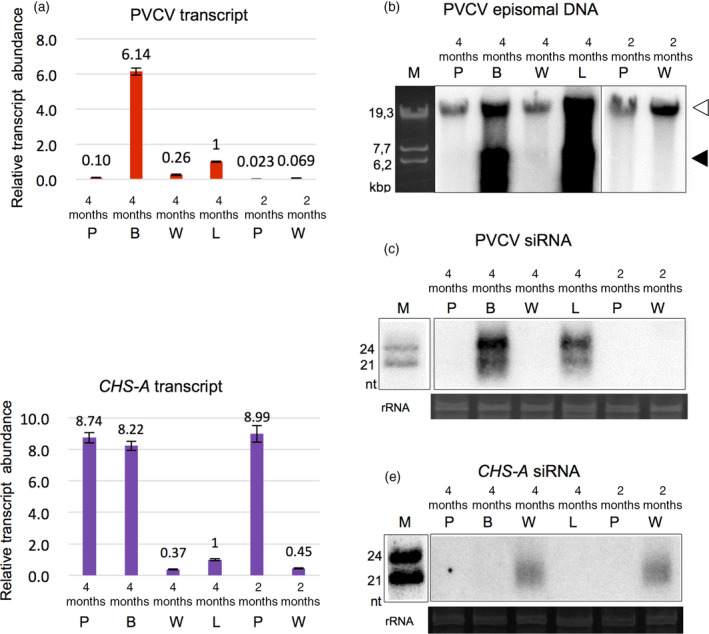
Figure 4.

Activation of petunia vein clearing caulimovirus (PVCV) in the blotched region of petals. DNA and total RNAs were isolated from pigmented (4m P), white (4m W) and blotched (4m B) regions of petals and leaves with the vein‐clearing symptom (4m L) of 4‐month‐old plants, and pigmented (2m P) and white (2m W) regions of petals of 2‐month‐old plants. Relative abundance of PVCV transcripts (a) and CHS‐A transcripts (d) was determined by qPCR and normalized to the abundance of Actin7 transcripts. Error bars indicate the standard errors of three biological replications. (b) Proviral (white arrowhead) and episomal (black arrowhead) PVCV DNAs were detected by Southern blot hybridization. PVCV (c) and CHS‐A (e) siRNAs were detected by northern blot hybridization. Ribosomal RNAs stained by ethidium bromide are shown as loading controls in (c) and (e). Molecular weight markers of linear dsDNAs (b) and ssRNAs (c and e) are indicated as lane M.
