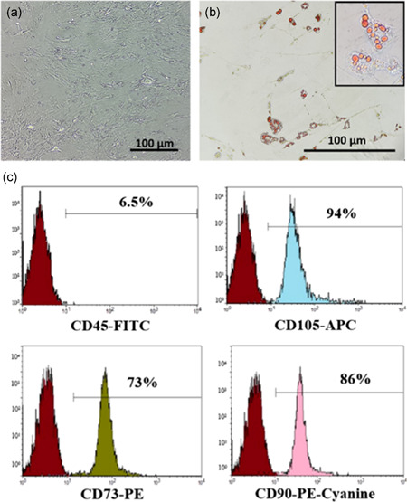
Figure 1.

Characterization of adipose tissue‐derived mesenchymal stem cells (AD‐MSCs). Representative images and flow plots of (a) spindle shape of AD‐MSCs after 7 days as determined using phase‐contrast microscopy (×200). (b) Adipogenic differentiation after 21 days was determined by Oil Red O staining (×200). (c) Immunophenotyping of Passage 2 AD‐MSCs using flow cytometric analysis of CD45, CD105, CD73, and CD90 staining
