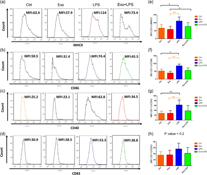
Figure 5.

The effect of mesenchymal stromal cell‐derived exosomes and/or lipopolysaccharide (LPS) on dendritic cell (DC) expression of surface markers. The phenotype of immature DCs (Ctrl), MDC‐derived exosome (Exo)‐treated DCs, LPS‐treated DCs and Exo + LPS‐treated DCs were assessed by flow cytometry. DC cells were gated by CD11c+ MHCII+, and then based on this gate, a histogram was drawn up. Representative histogram shows the expression of MHCII (a). DC cells were gated by CD11c+CD86+, and then based on this gate, a histogram was drawn up. Representative histogram shows the expression of CD86 (b). DC cells were gated by CD11c+CD40+, and then based on this gate, a histogram was drawn up. Representative histogram shows the expression of CD40 (c). DC cells were gated by CD11c+CD83+, and then based on this gate, a histogram was drawn up. Representative histogram shows the expression of CD83 (d). Bar graphs represent mean fluorescence intensity (MFI; arbitrary units [AU] of fluorescence) ± SEM percentage expression of CD11c‐MHCII (e) and costimulatory molecules CD86 (f), CD40 (g), and CD83 (h) in the various groups from n = 3 independent experiments. *p < .05, **p < .01. SEM, standard error of the mean
