Figure 2.

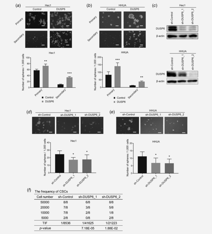
DUSP6 promotes sphere formation and self‐renewal in endometrial cancer cells. (a) Bright‐phase images of primary and secondary spheres formed by DUSP6‐overexpressing Hec1 cells. Scale bar, 100 μm. The bar graph shows the number of spheres/1,000 cells that were ≥50 μm in diameter. (b) Bright‐phase images of primary and secondary spheres formed by DUSP6‐overexpressing HHUA cells. Scale bar, 100 μm. The bar graph shows the number of spheres/1,000 cells that were ≥50 μm. (c) Human DUSP6 was knocked down by shRNA (sh‐DUSP6) in Hec1 and HHUA cells. The protein level of DUSP6 was measured by Western blot analysis. β‐Actin was also measured as a loading control. (d) Bright‐phase images of spheres formed by DUSP6‐knockdown Hec1 cells. Scale bar, 100 μm. The bar graph shows the number of spheres/1,000 cells that were ≥50 μm. (e) Bright‐phase images of spheres formed by DUSP6‐knockdown HHUA cells. Scale bar, 100 μm. The bar graph shows the number of spheres/1,000 cells that were ≥50 μm. (f) In vivo limiting dilution assay showing the TIF of DUSP6‐knockdown Hec1 cells. The TIF was calculated using ELDA software. Data are representative of at least three independent experiments. Error bars indicate the standard deviation. *p < 0.05, **p < 0.01, ***p < 0.005.
