Figure 4.

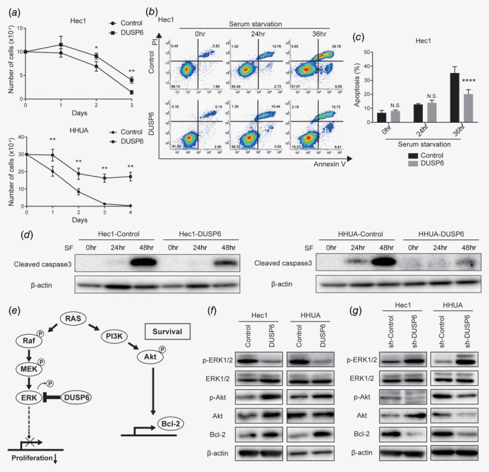
DUSP6 promotes resistance to serum starvation and inhibits apoptosis via Akt phosphorylation, potentially contributing to acquisition of a CSC phenotype. (a) Growth curves of DUSP6‐overexpressing Hec1 and HHUA cells cultured in serum‐free DMEM. Cells were counted daily. (b) Evaluation of apoptosis by flow cytometric analysis of annexin V in control and DUSP6‐overexpressing Hec1 cells cultured in serum‐free DMEM for the indicated number of days. The annexin V‐positive cells are gated on the right. The numbers indicate the population of each gate. (c) Bar graph showing the proportions of annexin V‐positive cells. (d) Western blot analysis of cleaved caspase 3 in control and DUSP6‐overexpressing Hec1 and HHUA cells. The cells were cultivated in serum‐free DMEM for 24 or 48 hr. β‐actin was also measured as a loading control. (e) Schematic of the signaling pathways that activate Akt phosphorylation and Bcl‐2, and the cross‐regulation between the Akt and MAPK signaling pathways. (f) Western blot analyses of p‐ERK1/2, ERK1/2, p‐Akt, Akt and Bcl‐2 in control and DUSP6‐overexpressing Hec1 and HHUA cells. β‐Actin was also measured to ensure equal loading of a gel and a representative loading control for all sample is shown. (g) Western blot analyses of p‐ERK1/2, ERK, p‐Akt, Akt and Bcl‐2 in sh‐Control and DUSP6‐knockdown Hec1 and HHUA cells. β‐Actin was also measured to ensure equal loading of a gel and a representative loading control for all sample is shown. Data are representative of at least three independent experiments. Error bars indicate standard deviations. *p < 0.05, **p < 0.01, ****p < 0.001, N.S., not significant.
