
Fig. 7.
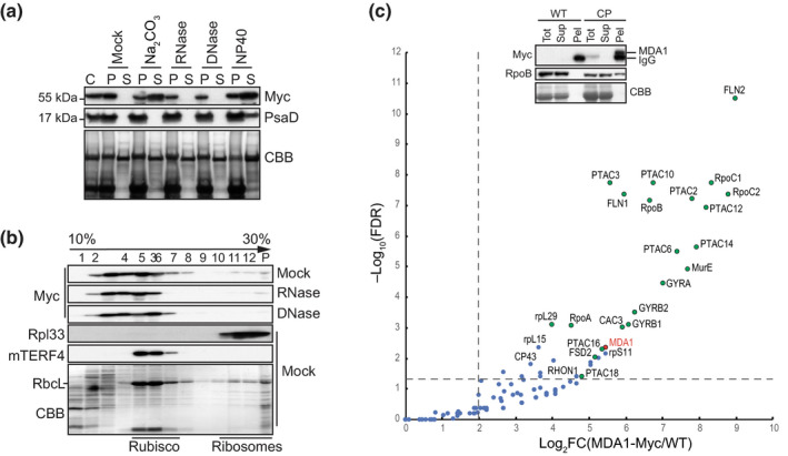
Arabidopsis MDA1 is found in high molecular weight (MW) complexes in vivo and associates with plastid‐encoded RNA polymerase (PEP) and transcriptional active chromosome (TAC) components. (a) MDA1 is a peripheral membrane protein whose membrane association is RNA and DNA‐independent. Chloroplast membrane fractions (C) were mock‐treated or 0.2 M Na2CO3, RNase, DNase, 1% NP40 treated before centrifugation. The presence of MDA1 in the pellet (P) or soluble fraction (S) after treatment was analyzed by immunoblotting with an antibody against the Myc epitope or the chloroplast integral membrane protein, PsaD as a control. The membrane stained with Coomassie Blue (CBB) is shown below. (b) Sucrose gradient fractionation of mock, RNase‐ and DNase‐treated chloroplast extracts. An equal volume of each fraction was analyzed on immunoblots using antibodies indicated at left. The chloroplast RNA intron splicing factor proteins, mTERF4 and the ribosomal protein Rpl33 are found in high MW complexes of c. 0.55 MDa and 1 MDa, respectively (Hammani & Barkan, 2014) and serve as size fractionation controls. (c) Chloroplast protein interactome of MDA1. MDA1 co‐immunoprecipitates (coIP) with components of the plastid TAC (Pfalz et al., 2006) including the subunits of the plastid encoded RNA polymerase. Solubilized chloroplasts from complemented mda1 plants expressing the 4Myc‐tagged MDA1 (CP) or wild‐type (WT) plants were used for immunoprecipitation with anti‐Myc antibody and coIP proteins were identified by MS analysis. Volcano plots show the enrichment of proteins copurified with 4Myc‐tagged MDA1 as compared with control IPs. IPs were performed on biological triplicate. The y‐ and x‐axes display the negative common logarithm of the adjusted false discovery rate (FDR) and fold‐changes, respectively. The dashed lines indicate the threshold above which proteins are significantly enriched (FDR < 0.05 and FC > 4). TAC components (Pfalz et al., 2006) are represented as green dots. The immunoblot validation of RpoB association is shown. Replicate immunoblots were probed with anti‐Myc or RpoB antibody. The full list of MDA1‐associated proteins is available in Supporting Information Table S4.