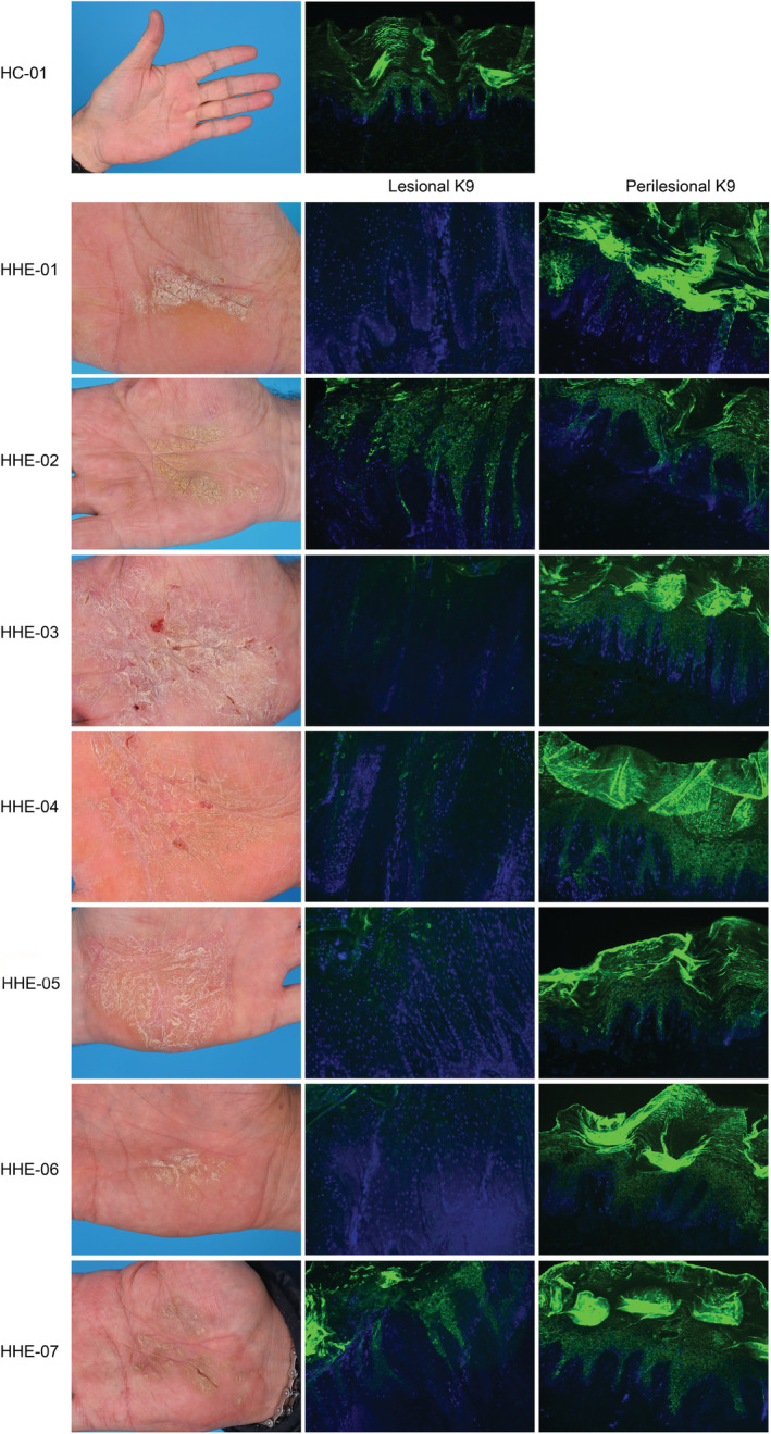
FIGURE 1.

In the left column, clinical pictures showing the palms of the seven patients with a clinical phenotype of hyperkeratotic hand eczema. The two columns at the right show the K9 immunofluorescence staining patterns of their skin biopsies: reduced or absent staining of keratin 9 in lesional skin (5/7, middle column) and normal K9 staining in perilesional (7/7, right column) and healthy control (HC) skin
