Fig. 4.

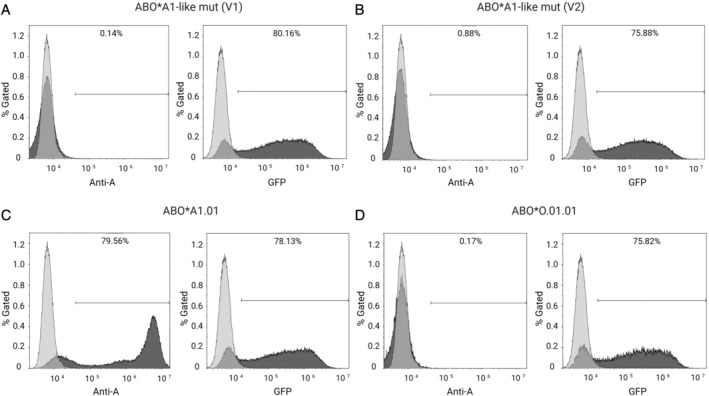
Representative flow cytometry analyses of transfected HeLa cells for blood group A antigen and GFP expression. The used anti‐human blood group A monoclonal antibody binds specifically to blood group A expressed on transfected HeLa cells. A or GFP expression is indicated (dark gray filled) and untreated HeLa cells were used as control cells (light gray, respective control). (A) ABO*A1‐like mut (V1) transfected cells with 80% GFP expression, indicating positive transfection. (B) ABO*A1‐like mut (V2) transfected cells with 75% GFP expression. Despite good transfection efficiencies, V1 and V2 did not result in expression of A antigen. (C) Positive control, ABO*A1.01 (wild type) transfectants, indicating 79% A and 78% GFP expression. (D) Negative control, ABO*O.01.01 transfectants, demonstrating a lack of A expression but indicating 75% of GFP expressing cells.
