Figure 4.

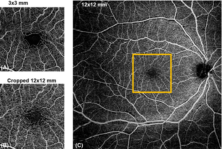
Comparison of A) 3 × 3‐mm2 scan, B) cropped 12 × 12‐mm2 scan and C) original 12 × 12‐mm2 scan of the superficial vascular plexus. A 3 × 3‐mm2 area centred on the foveal avascular zone was selected on 12 × 12‐mm2 scan (orange box).

Comparison of A) 3 × 3‐mm2 scan, B) cropped 12 × 12‐mm2 scan and C) original 12 × 12‐mm2 scan of the superficial vascular plexus. A 3 × 3‐mm2 area centred on the foveal avascular zone was selected on 12 × 12‐mm2 scan (orange box).