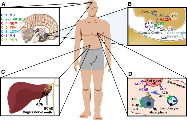
Fig. 1.

The cholinergic system. (A) The human brain includes eight cholinergic nuclei. Ch1 in the medial septum, Ch2 and Ch3 in the vertical and horizontal limbs of the diagonal band of Broca, Ch4 in the nucleus basalis of Meynert, Ch5 in the pedunculopontine nucleus, Ch6 in the laterodorsal tegmental nucleus, Ch7 in the medial habenula, and Ch8 in the para‐bigeminal nucleus [12]. (B) ChAT in the presynaptic cell synthesizes ACh from choline and acetyl‐CoA. VAChT packages ACh in vesicles, which are secreted to the cleft. There, ACh can activate pre (auto)‐ and postsynaptic cholinergic receptors (nicotinic or muscarinic). ACh in the cleft is hydrolyzed to acetate and choline by acetylcholinesterase (AChE) which is attached to the cellular membrane by proline‐rich membrane anchor 1 (PRIMA1, in the brain) or Acetylcholinesterase collagenic tail Q peptide (ColQ, in neuromuscular junctions). Choline transporters (CHT) transporters reuptake choline from the cleft to the presynaptic cell [1, 13]. (C) The vagus nerve reaches internal organs such as the liver, where it intercepts information and attenuates inflammation via ACh blockade of the NFkB pathway. In the liver, the main cholinesterase enzyme is butyrylcholinesterase (BChE) [13, 14]. (D) In the blood, ACh secreted by immune cells such as lymphocytes is intercepted by the α7 nicotinic receptor of other immune cells (e.g., macrophages), which reduces their inflammatory signal [TNF, interleukin (IL)‐1β, IL‐6]. Blood ACh can be hydrolyzed by AChE on the membrane of red blood cells [13].
