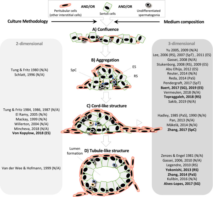
Figure 1.

Structural reorganization of all or combinations of testicular cell types in‐vitro in chronological order. Testicular tubulogenesis in‐vitro comprises distinct phases: gaining of cell confluence (A). Aggregation of Sertoli cells into multinodular mounds under influence of contractile peritubular myoid cells (B). Interconnection and merging of multinodular mounds to form cable‐like structures (C). Formation of hollow tubules (D). A shift from 2D (light gray table) toward 3D (dark gray table) culture models has been observed because of the latter's ability to improve the cell reorganization. Aside from the culture methodology, medium composition influences the different aspects of testicular tubulogenesis in‐vitro. Of the different media‐ingredients, KSR has been proven critical (bold). Upon reorganization of the testicular cells, differentiation of spermatogonia could be seen. The most advanced differentiation stage in each study was indicated between parentheses. ES, elongates spermatids; PaS, pachytene spermatocytes; RS, round spermatids; SG, spermatogonia; SpC, spermatocytes; SpT, spermatids
