
Figure 1.
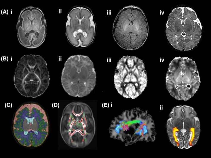
(A) (i) T1‐ and (ii) T2‐weighted images of an infant at 26 weeks gestational age (GA) and (iii) T1‐ and (iv) T2‐weighted images of an infant at 42 weeks GA at the level of the basal ganglia. (B) Diffusion magnetic resonance imaging maps at the level of the basal ganglia (i) fractional anisotropy, FA (ii) mean diffusivity, MD (iii) orientation dispersion index, ODI and (iv) neurite density index, NDI. (C) Brain segmentation in an infant born at 27+4 weeks gestational age and imaged at 41+2 weeks postmenstrual age. Key: Green = cortical grey matter, blue = white matter, grey = deep grey matter, pink = extracerebral cerebrospinal fluid. (D) Correlation between gestational age at birth and FA measures in white matter assessed using tract‐based spatial statistics. Mean FA skeleton (green) overlaid on mean FA map in the axial plane. Voxels showing a significant correlation (P < 0.05) between GA at birth FA are shown in red. (E) Diffusion MR tractography (i) arcuate fasciculus and (ii) optic radiations