
Figure 2.
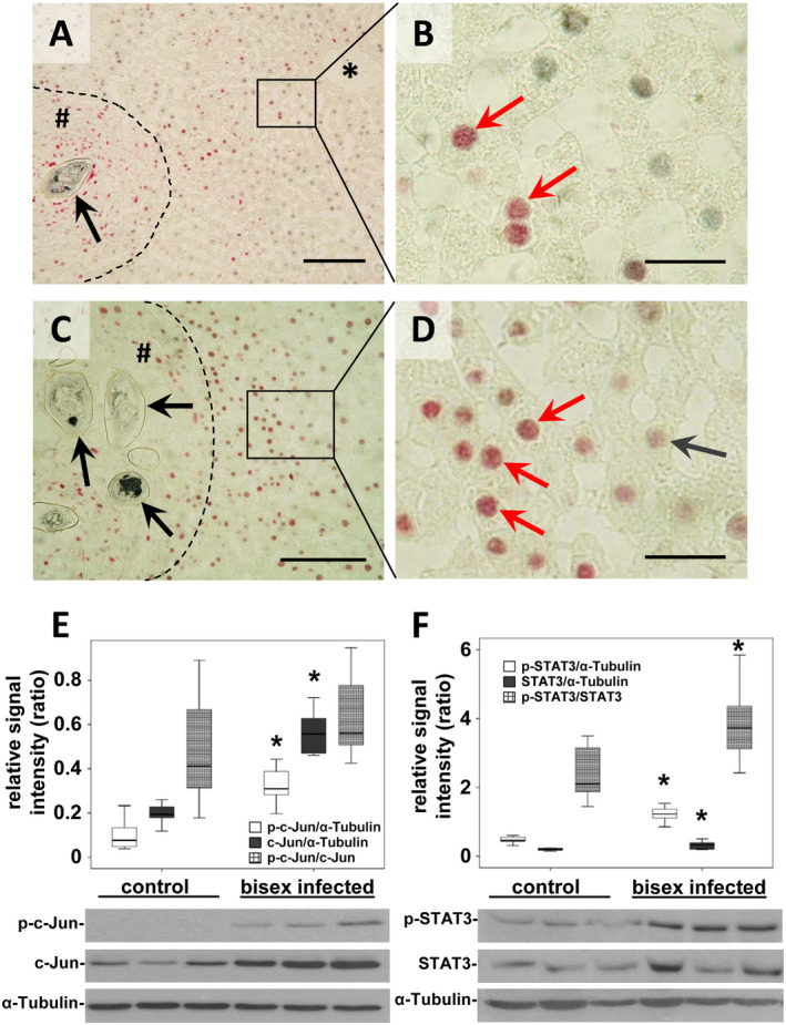
Activation of c‐Jun in hepatocytes around S. mansoni egg–induced granuloma. (A‐D) Coimmunostaining (red arrows) for c‐Jun (red) and HNF4α (gray) depicted the nuclear translocation of c‐Jun in hepatocytes joining granuloma and central veins (*). Note the directional hepatocellular activation pattern of c‐Jun from granuloma to central vein (A). Enhanced nuclear translocation of c‐Jun (red arrows) next to granuloma around egg‐conglomerates (C,D) *central vein, # granuloma, dashed line granuloma border, black arrow S. mansoni egg. Magnification ×200 (A), ×1,000 (B,D), ×400 (C), bars 100 µm (A,C), 20 µm (B,D). (E) Semiquantitative western blot analysis and subsequent assessment of optical density revealed enhanced expression and activation of c‐Jun (p‐Ser73) in S. mansoni (bisex infected, ♀+♂)–infected hamsters in comparison with single‐sex infected hamsters (no egg production) that served as controls. Note that the ratio of p‐c‐Jun/c‐Jun increased only by trend. (F) Western blot analysis demonstrated induced expression and activation of STAT3 (p‐Ser705) in S. mansoni (♀+♂)–infected hamsters in comparison with controls. n = 9/group for statistical analysis; representative blots are depicted, *P < 0.05.